PET Scan for Merkel Cell Carcinoma: Diagnosis and Staging
16 May, 2023
 Obaidullah Junaid
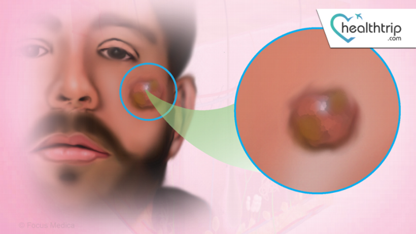
Merkel cell carcinoma (MCC) is a rare and aggressive form of skin cancer that develops in the Merkel cells located in the skin's basal layer. Merkel cells are responsible for detecting touch and pressure in the skin. MCC is usually found in sun-exposed areas of the skin and is more common in older adults and people with weakened immune systems. PET scan is an imaging technique that can be used to diagnose and stage MCC.
PET scan stands for Positron Emission Tomography. It is an imaging technique that uses a radioactive tracer to produce images of the body's metabolic activity. The tracer is injected into the patient's vein, and the PET scanner detects the gamma rays emitted by the tracer as it travels through the body. The images produced by the PET scan show areas of high metabolic activity, which can indicate the presence of cancer cells.
Transform Your Beauty, Boost Your Confidence
Find the right cosmetic procedure for your needs.
We specialize in a wide range of cosmetic procedures

PET scans are particularly useful for diagnosing and staging MCC because this cancer has a high rate of metastasis. MCC often spreads to nearby lymph nodes and distant organs such as the lungs, liver, and brain. PET scans can detect these metastases and provide information on the extent of the cancer's spread.
Diagnosis of MCC using PET scan:
The diagnosis of MCC typically involves a biopsy, in which a small sample of the tumor is removed and examined under a microscope. However, PET scans can also be used to diagnose MCC, particularly in cases where the tumor is difficult to biopsy, or there is a suspicion that the cancer has spread beyond the primary tumor.
PET scans are particularly useful for diagnosing MCC because they can detect areas of high metabolic activity that may indicate the presence of cancer cells. In MCC, these areas of high metabolic activity are usually found in the primary tumor site and nearby lymph nodes.
To perform a PET scan, the patient is injected with a radioactive tracer, which is typically FDG (fluorodeoxyglucose). FDG is a radioactive form of glucose that is taken up by cells in the body, particularly cancer cells, which have a higher rate of glucose metabolism than normal cells. The tracer takes about an hour to circulate through the body, and the patient is then positioned in the PET scanner.
The scanner detects the gamma rays emitted by the tracer and produces images of the body's metabolic activity. Areas of high metabolic activity appear as "hot spots" on the images. The PET scan can detect the primary tumor site and any areas of metastasis, such as lymph nodes or distant organs.
Most popular procedures in India
Total Hip Replacemen
Upto 80% off
90% Rated
Satisfactory

Total Hip Replacemen
Upto 80% off
90% Rated
Satisfactory

Total Hip Replacemen
Upto 80% off
90% Rated
Satisfactory

ANGIOGRAM
Upto 80% off
90% Rated
Satisfactory

ASD Closure
Upto 80% off
90% Rated
Satisfactory

Staging of MCC using PET scan:
Staging is the process of determining the extent of cancer spread. In MCC, staging is particularly important because the cancer has a high rate of metastasis. Accurate staging is crucial for determining the best treatment approach and predicting the patient's prognosis.
PET scans are an important tool for staging MCC because they can detect areas of metastasis that may not be visible on other imaging tests. PET scans can detect metastases in lymph nodes, distant organs, and bones.
PET scans are often used in conjunction with other imaging tests, such as CT scans and MRI scans, to provide a comprehensive picture of the cancer's spread. CT scans can provide detailed images of the internal organs and bones, while MRI scans can provide high-resolution images of the brain and soft tissues.
The combination of PET, CT, and MRI scans can provide a detailed map of the cancer's spread, which is essential for accurate staging and treatment planning.
Treatment of MCC:
The treatment of MCC depends on the stage of the cancer and the patient's overall health. Treatment options include surgery, radiation therapy, chemotherapy, and immunotherapy.
Surgery is often the first line of treatment for early-stage MCC. The goal of surgery is to remove the primary tumor and any nearby lymph nodes that may contain cancer cells. Surgery is often followed by radiation therapy to kill any remaining cancer cells and reduce the risk of recurrence.
In more advanced cases of MCC, radiation therapy may be used as the primary treatment or in combination with chemotherapy. Radiation therapy uses high-energy radiation to kill cancer cells. Chemotherapy involves the use of drugs to kill cancer cells. Immunotherapy is a newer type of treatment that uses the body's own immune system to attack cancer cells.
PET scans are useful for monitoring the response to treatment. After treatment, a PET scan can be used to determine if the cancer has been successfully treated or if there are any remaining cancer cells. PET scans can also be used to detect recurrence of the cancer.
Limitations of PET scans for MCC:
While PET scans are useful for diagnosing and staging MCC, they do have limitations. PET scans cannot distinguish between cancer cells and other types of cells that have high metabolic activity, such as inflammation. This can lead to false-positive results, where areas of high metabolic activity are mistaken for cancer when they are actually caused by inflammation or infection.
PET scans also have limited spatial resolution compared to other imaging techniques such as CT scans and MRI scans. This means that PET scans may not be able to detect small tumors or small areas of metastasis. Therefore, PET scans are often used in combination with other imaging techniques to provide a more comprehensive picture of the cancer's spread.
Conclusion
PET scans are a useful tool for diagnosing and staging Merkel cell carcinoma. MCC is a rare and aggressive form of skin cancer that has a high rate of metastasis. PET scans can detect areas of high metabolic activity that may indicate the presence of cancer cells. PET scans can also detect areas of metastasis in lymph nodes, distant organs, and bones.
Accurate staging is crucial for determining the best treatment approach and predicting the patient's prognosis. PET scans are often used in conjunction with other imaging tests, such as CT scans and MRI scans, to provide a comprehensive picture of the cancer's spread.
While PET scans have limitations, they are a valuable tool for monitoring the response to treatment and detecting recurrence of the cancer. Overall, PET scans play an important role in the diagnosis, staging, and treatment of Merkel cell carcinoma.
Wellness Treatment
Give yourself the time to relax
Lowest Prices Guaranteed!

Lowest Prices Guaranteed!