
Understanding the Different Types of Imaging Tests
16 Apr, 2023
Medical imaging plays a critical role in modern healthcare, allowing doctors to visualize the internal structures of the human body without invasive procedures. Imaging tests are used for various purposes, from diagnosing diseases to monitoring treatment progress. With advances in technology, there are now several different types of imaging tests available, each with its own unique features and applications. In this blog, we will explore some of the most common types of imaging tests used in medical practice, their principles, and their specific uses.
- X-ray Imaging
X-ray imaging is one of the oldest and most widely used types of imaging tests. It involves passing a small amount of ionizing radiation through the body, which is absorbed by different tissues to varying degrees, creating an image on a detector. X-ray images are typically used to visualize bones and can help detect fractures, infections, tumors, and other abnormalities in the skeletal system. X-rays are also used in dentistry to evaluate teeth and jaw structure.
Transform Your Beauty, Boost Your Confidence
Find the right cosmetic procedure for your needs.
We specialize in a wide range of cosmetic procedures

One of the advantages of X-ray imaging is its quick and relatively low-cost nature. However, it does have some limitations. X-rays are not effective at visualizing soft tissues such as organs or blood vessels, and the ionizing radiation used in X-rays can be harmful in high doses. Therefore, X-rays are generally used sparingly and with caution, particularly in pregnant women and children.
- Computed Tomography (CT) Scan
Computed Tomography, commonly known as CT scan, is a more advanced imaging test that uses X-ray technology to create detailed cross-sectional images of the body. CT scans provide a 3D view of the internal organs, bones, blood vessels, and soft tissues, making it a valuable tool in diagnosing a wide range of conditions, including cancer, vascular diseases, and trauma injuries.
CT scans work by rotating an X-ray tube and a detector around the body, capturing multiple images from different angles. These images are then processed by a computer to create detailed cross-sectional slices that can be reconstructed into 3D images. CT scans are particularly useful in detecting small lesions or abnormalities that may not be visible on X-rays.
One of the drawbacks of CT scans is that they involve a higher dose of ionizing radiation compared to X-rays, which can increase the risk of radiation exposure, particularly in repetitive or multiple scans. However, modern CT scanners are equipped with dose-reduction techniques to minimize radiation exposure, and the benefits of CT scans often outweigh the risks in many clinical situations.
- Magnetic Resonance Imaging (MRI)
Magnetic Resonance Imaging, commonly known as MRI, is a non-invasive imaging test that uses strong magnets and radio waves to create detailed images of the body's internal structures. MRI provides excellent visualization of soft tissues such as organs, muscles, nerves, and blood vessels, making it a valuable tool in diagnosing conditions such as tumors, joint injuries, and neurological disorders.
Unlike X-rays and CT scans, MRI does not involve ionizing radiation, making it a safer option for certain populations, including pregnant women and children. However, MRI has its own limitations. It may not be suitable for patients with certain metallic implants, as the strong magnetic field can interfere with the function of the implant or cause injury. Additionally, MRI scans are generally more time-consuming and expensive compared to other imaging tests.
Most popular procedures in
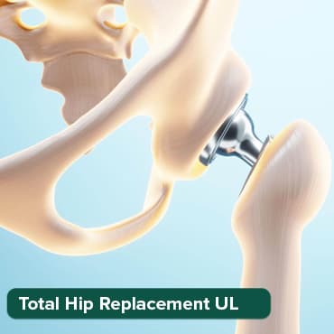
Total Hip Replacemen
Upto 80% off
90% Rated
Satisfactory
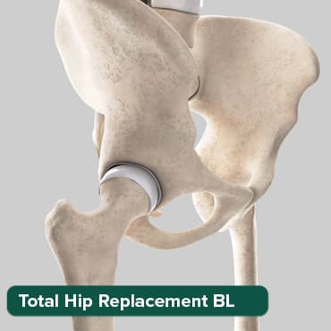
Total Hip Replacemen
Upto 80% off
90% Rated
Satisfactory
Breast Cancer Surger
Upto 80% off
90% Rated
Satisfactory

Total Knee Replaceme
Upto 80% off
90% Rated
Satisfactory
Total Knee Replaceme
Upto 80% off
90% Rated
Satisfactory
- Ultrasound
Ultrasound, also known as sonography, is a type of imaging test that uses high-frequency sound waves to create real-time images of the body's internal structures. Ultrasound is commonly used to visualize organs such as the heart, liver, kidneys, and reproductive organs, as well as to monitor fetal development during pregnancy.
Ultrasound works by transmitting sound waves into the body through a handheld device called a transducer, which also receives the echoes continue
of the sound waves as they bounce back from the tissues. These echoes are then processed by a computer to create images that can be viewed in real-time. Ultrasound is safe, non-invasive, and does not involve any ionizing radiation, making it suitable for a wide range of patients, including pregnant women and children.
One of the unique features of ultrasound is its ability to provide dynamic images, allowing doctors to assess the movement and function of organs in real-time. This makes it valuable in assessing conditions such as heart function, blood flow, and fetal development. Ultrasound is also commonly used for guiding procedures such as biopsies and drainages, as it provides real-time visualization of the needle and surrounding structures.
- Nuclear Medicine Imaging
Nuclear medicine imaging is a specialized type of imaging that involves the use of radioactive materials to visualize the function and metabolism of organs and tissues in the body. In nuclear medicine, a small amount of radioactive material, called a radiotracer, is administered to the patient either orally, intravenously, or by inhalation. The radiotracer accumulates in the target organ or tissue and emits gamma rays, which are detected by a gamma camera or a PET (Positron Emission Tomography) scanner to create images.
Nuclear medicine imaging is commonly used to evaluate organ function, such as the heart, lungs, thyroid, and bones. It is also used in cancer imaging, as some radiotracers are specifically designed to accumulate in cancer cells, helping in cancer detection, staging, and monitoring treatment response.
One of the advantages of nuclear medicine imaging is its ability to provide functional information, showing how organs and tissues are functioning rather than just their anatomical structure. However, it does involve the use of radioactive materials, which can expose patients to ionizing radiation. The benefit of the information obtained from nuclear medicine imaging is carefully weighed against the risks of radiation exposure, and strict safety protocols are followed to minimize radiation exposure to patients and healthcare providers.
- Interventional Radiology
Interventional radiology is a specialized field of radiology that involves performing minimally invasive procedures using imaging guidance. Interventional radiologists use imaging techniques such as X-ray, CT scan, MRI, and ultrasound to visualize the target area and guide the placement of catheters, needles, or other instruments to diagnose and treat various conditions. Interventional radiology procedures can include angiography, embolization, biopsies, drainages, and tumor ablation, among others.
Interventional radiology procedures are often less invasive and have shorter recovery times compared to traditional surgical procedures, making them a preferred option for certain conditions. They also carry fewer risks and complications compared to open surgery, as they do not involve large incisions. Interventional radiology has revolutionized the field of medicine, allowing for precise and targeted treatments without the need for major surgery.
In conclusion, imaging tests are invaluable tools in modern healthcare for diagnosing, monitoring, and treating various conditions. They provide visual information about the internal structures of the body, helping doctors make accurate diagnoses and treatment plans. From X-ray imaging to CT scans, MRI, ultrasound, nuclear medicine imaging, and interventional radiology, each type of imaging test has its own unique features, benefits, and limitations. The choice of imaging test depends on the specific condition being evaluated, the patient's age, medical history, and other factors, and is determined by the physician based on the best available evidence and clinical judgment. It's important to discuss any concerns or questions about imaging tests with your healthcare provider to ensure that you have a clear understanding of the purpose, benefits, and risks of the test. With advancements in imaging technology, these tests continue to evolve and play a crucial role in improving patient care and outcomes.
Additionally, it's essential to understand that imaging tests are just one piece of the puzzle in medical diagnosis and treatment. They are often used in conjunction with other clinical assessments, such as patient history, physical examinations, laboratory tests, and other diagnostic procedures, to form a comprehensive understanding of a patient's condition.
Wellness Treatment
Give yourself the time to relax
Lowest Prices Guaranteed!

Lowest Prices Guaranteed!