PET Scan for Thymoma: Diagnosis and Staging
16 May, 2023
Thymoma is a rare type of cancer that originates in the thymus gland, which is located in the upper chest, just behind the breastbone. The thymus gland plays a crucial role in the immune system, and thymoma can cause a variety of symptoms, including chest pain, shortness of breath, and coughing. While thymoma can be difficult to diagnose and treat, advances in medical imaging technology have made it easier to identify and stage the disease. One such technology is the PET scan, which uses radioactive tracers to create detailed images of the body’s internal structures.
In this blog, we’ll take a closer look at thymoma, its symptoms and diagnosis, and the role that PET scans play in staging the disease.
Transform Your Beauty, Boost Your Confidence
Find the right cosmetic procedure for your needs.
We specialize in a wide range of cosmetic procedures

Thymoma: Symptoms and Diagnosis
Thymoma is a rare type of cancer, accounting for only 0.2 to 1.5% of all malignancies. It is more common in men than women, and typically presents in individuals over the age of 40. Thymoma is often asymptomatic, and is frequently discovered incidentally during medical imaging studies performed for other reasons. However, when thymoma does cause symptoms, they can be quite varied, and may include:
- Chest pain or discomfort
- Shortness of breath
- Coughing
- Difficulty swallowing
- Hoarseness
- Fatigue
- Weakness
- Fever
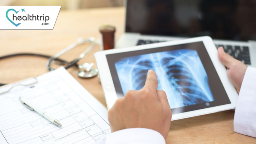
The diagnosis of thymoma typically involves a combination of medical history, physical examination, and imaging studies. Blood tests may also be used to look for elevated levels of certain proteins or markers associated with thymoma. Imaging studies such as chest X-rays, CT scans, and MRI scans can provide detailed information about the size, location, and characteristics of the tumor. In some cases, a biopsy may also be necessary to confirm the diagnosis.
PET Scans and Thymoma
Positron emission tomography (PET) is a medical imaging technique that uses radioactive tracers to create detailed images of the body’s internal structures. During a PET scan, a small amount of radioactive material is injected into the patient’s bloodstream. This material is absorbed by the body’s tissues and organs, and emits gamma rays that can be detected by a special camera. The camera creates images of the body based on the distribution of the radioactive material, allowing doctors to see how different tissues and organs are functioning.
PET scans can be used to diagnose and stage thymoma, as well as to monitor the effectiveness of treatment. PET scans are particularly useful for detecting small or early-stage tumors that may not be visible on other types of imaging studies. They can also help doctors determine whether the cancer has spread to other parts of the body, which is a critical factor in determining the most appropriate treatment approach.
PET scans are typically performed in combination with CT scans or MRI scans, which provide detailed anatomical information about the body. The PET and CT or MRI scans are fused together to create a comprehensive image of the patient’s anatomy and metabolic activity. This combined approach is known as PET/CT or PET/MRI imaging.
PET scans are particularly useful in diagnosing and staging thymoma because the radioactive tracers used in the scans tend to accumulate in areas of the body with high metabolic activity. Cancer cells typically have a higher metabolic rate than healthy cells, which means that they absorb more of the radioactive material and show up as bright spots on the PET scan. This can make it easier for doctors to identify the location and extent of the tumor.
Most popular procedures in India
Atrial septal defect
Upto 80% off
90% Rated
Satisfactory

Coronary Angiogram a
Upto 80% off
90% Rated
Satisfactory
Coronary Angiogram C
Upto 80% off
90% Rated
Satisfactory

Liver Transplant
Upto 80% off
90% Rated
Satisfactory
Total Hip Replacemen
Upto 80% off
90% Rated
Satisfactory

PET scans can also be useful in monitoring the effectiveness of treatment for thymoma. After treatment, PET scans can be used to check for residual tumor activity and to see whether the cancer has spread or recurred. This can help doctors determine whether additional treatment is necessary, and if so, what type of treatment would be most effective.
Staging Thymoma with PET Scans
Staging is the process of determining the extent and severity of cancer, which is critical in determining the most appropriate treatment approach. Staging for thymoma typically involves a combination of imaging studies, biopsy, and surgical exploration. PET scans can play an important role in this process, particularly in identifying metastases, or the spread of cancer to other parts of the body.
There are four stages of thymoma, ranging from Stage I (localized tumor without invasion of surrounding structures) to Stage IV (metastatic disease). The staging of thymoma is based on a number of factors, including the size and location of the tumor, the degree of invasion into surrounding structures, and the presence of metastases.
PET scans can help identify the presence and location of metastases, which is particularly important in determining the stage of the disease. In addition to detecting metastases, PET scans can also help identify areas of the tumor that are particularly active or aggressive, which can help guide treatment decisions.
Treatment Options for Thymoma
The treatment of thymoma depends on a variety of factors, including the stage and location of the tumor, the patient’s age and overall health, and the preferences of the patient and their healthcare team. Treatment options for thymoma may include surgery, radiation therapy, chemotherapy, or a combination of these approaches.
Surgery is the primary treatment for localized thymoma, and may involve removal of the thymus gland, as well as any surrounding tissue or lymph nodes that may be affected by the tumor. In some cases, surgery may be followed by radiation therapy to destroy any remaining cancer cells.
For more advanced or metastatic thymoma, chemotherapy may be used to help shrink the tumor and slow its growth. Radiation therapy may also be used to destroy cancer cells and alleviate symptoms.
In cases where thymoma has spread to other parts of the body, treatment may focus on managing symptoms and improving quality of life. This may involve palliative care, which aims to relieve pain and other symptoms of the disease.
Conclusion
Thymoma is a rare type of cancer that can be difficult to diagnose and treat. Advances in medical imaging technology, such as PET scans, have made it easier to identify and stage the disease, which is critical in determining the most appropriate treatment approach.
PET scans use radioactive tracers to create detailed images of the body’s internal structures, which can be particularly useful in detecting small or early-stage tumors, identifying areas of the tumor that are particularly active or aggressive, and monitoring the effectiveness of treatment. PET scans can also help identify the presence and location of metastases, which is critical in determining the stage of the disease and guiding treatment decisions.
While thymoma can be challenging to treat, there are a variety of treatment options available, including surgery, radiation therapy, chemotherapy, and palliative care. By working closely with their healthcare team and staying informed about the latest advances in medical technology, patients with thymoma can improve their chances of a successful outcome and achieve a better quality of life.
Wellness Treatment
Give yourself the time to relax
Lowest Prices Guaranteed!

Lowest Prices Guaranteed!