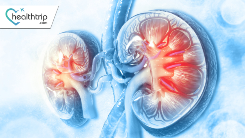
PET scan for Renal Cancer: Diagnosis and Staging

Book free consulting session with HealthTrip expert
Diagnosis of renal cancer typically involves a combination of medical history, physical examination, blood tests, imaging studies, and biopsy. PET scans are increasingly being used to diagnose and stage renal cancer, as they provide detailed information about the metabolic activity of the tumour and surrounding tissues.
In this blog, we will discuss how PET scans are used in the diagnosis and staging of renal cancer, their benefits, and potential risks.
How Does a PET Scan Work?
Before discussing the use of PET scans in renal cancer diagnosis, it is essential to understand how they work. PET scans involve the use of a small amount of radioactive material called a tracer, which is injected into the bloodstream. The tracer travels through the body and accumulates in areas of high metabolic activity, such as tumours.
Once the tracer has accumulated in the target tissue, a special camera detects the radiation emitted by the tracer and produces three-dimensional images of the tissue. These images can be used to assess the size, location, and metabolic activity of the tumour, as well as the spread of cancer to other parts of the body.
PET scan for Renal Cancer Diagnosis
Renal cancer can be challenging to diagnose, as it often has no symptoms in its early stages. When symptoms do occur, they can include blood in the urine, pain or discomfort in the side or lower back, or a lump or mass in the abdomen.
To diagnose renal cancer, healthcare providers typically begin with a physical exam and medical history. Blood and urine tests may be used to detect the presence of substances produced by kidney cancer cells. Imaging studies, such as ultrasound, CT scan, or MRI, may be used to visualise the tumour and surrounding tissues.
PET scans are increasingly being used to diagnose renal cancer, especially in cases where the tumour is difficult to visualise with other imaging techniques. PET scans can detect the metabolic activity of cancer cells, which may be higher than that of normal cells.
A study published in the Journal of Nuclear Medicine found that PET scans are more accurate in detecting renal cancer than CT scans. The study found that PET scans detected 93.1% of renal tumours, compared to 77.8% detected by CT scans.
PET scan for Renal Cancer Staging
Staging refers to the process of determining the extent and spread of cancer. Accurate staging is essential for determining the most appropriate treatment approach and predicting the prognosis of the disease.
Staging of renal cancer typically involves a combination of imaging studies, including CT scans, MRI, and PET scans. PET scans are increasingly being used in renal cancer staging, as they provide detailed information about the metabolic activity of the tumour and surrounding tissues.
PET scans can detect the presence of cancer cells in other parts of the body, such as the lymph nodes, bones, or lungs. This is important because renal cancer can spread to other parts of the body, even in its early stages.
A study published in the Journal of Urology found that PET scans were highly accurate in detecting the spread of renal cancer to other parts of the body. The study found that PET scans had a sensitivity of 92% and a specificity of 93%, meaning they were highly accurate
Benefits of PET Scans for Renal Cancer Diagnosis and Staging
PET scans have several benefits for the diagnosis and staging of renal cancer. These include:
1. Increased Accuracy: PET scans are more accurate than other imaging techniques in detecting renal cancer, especially in cases where the tumour is difficult to visualise with other imaging techniques.
2. Early Detection: PET scans can detect renal cancer in its early stages, before it spreads to other parts of the body.
3. Better Staging: PET scans provide detailed information about the metabolic activity of the tumour and surrounding tissues, allowing for more accurate staging of the cancer.
4. Treatment Planning: PET scans can help healthcare providers determine the most appropriate treatment approach based on the size, location, and spread of the tumour.
5. Monitoring: PET scans can be used to monitor the response of renal cancer to treatment over time.
Potential Risks of PET Scans
While PET scans are generally considered safe, they do involve exposure to a small amount of radiation. However, the amount of radiation exposure is typically less than that of a CT scan or X-ray. Additionally, some people may have an allergic reaction to the tracer used in the PET scan. Symptoms of an allergic reaction can include hives, itching, or difficulty breathing.
Preparing for a PET scan for Renal Cancer
Preparing for a PET scan for renal cancer typically involves
following some instructions provided by the healthcare provider. These instructions may include:
1. Fasting: Depending on the type of tracer used, patients may need to fast for several hours before the scan.
2. Medications: Patients may need to stop taking certain medications before the scan, as they may interfere with the results.
3. Rest: Patients may be advised to rest before the scan to reduce the risk of motion artefacts in the images.
4. Clothing: Patients should wear comfortable, loose-fitting clothing to the scan. Metal objects, such as jewellery or clothing with metal zippers, should be removed.
5. Hydration: Patients should drink plenty of water before and after the scan to help flush the tracer out of the body.
Conclusion
PET scans are increasingly being used in the diagnosis and staging of renal cancer. They provide detailed information about the metabolic activity of the tumour and surrounding tissues, allowing for more accurate diagnosis and staging. PET scans can also help healthcare providers determine the most appropriate treatment approach based on the size, location, and spread of the tumour. While PET scans are generally considered safe, they do involve exposure to a small amount of radiation. Patients should follow the instructions provided by their healthcare provider to prepare for the scan and minimise any potential risks.


