
PET Scan for Pancreatic Neuroendocrine Tumor: Diagnosis and Staging
16 May, 2023
Pancreatic neuroendocrine tumors (PNETs) are a rare type of pancreatic cancer that arises from the cells of the neuroendocrine system. While PNETs can be classified as either functional or non-functional, they are typically slow-growing and may not present with any symptoms until they have already spread to other parts of the body. Early detection and accurate staging of PNETs are essential for effective treatment and improved patient outcomes. One imaging modality that has proven to be highly effective in diagnosing and staging PNETs is the positron emission tomography (PET) scan.
PET Scan: How Does it Work?
Transform Your Beauty, Boost Your Confidence
Find the right cosmetic procedure for your needs.
We specialize in a wide range of cosmetic procedures

PET is a type of nuclear medicine imaging that uses a radioactive tracer to visualize metabolic activity in the body. The tracer is typically a small amount of a radioactive substance that is injected into the patient’s bloodstream. The tracer accumulates in cells that are actively metabolizing glucose, which is the primary source of energy for most cells in the body. The radioactive tracer emits positrons, which collide with electrons in the body and produce gamma rays. The gamma rays are detected by a PET scanner, which produces a 3D image of the distribution of the tracer in the body.
PET Scan for PNET Diagnosis
The diagnosis of PNETs can be challenging because they often do not present with any symptoms until they have already spread to other parts of the body. Additionally, PNETs can be difficult to distinguish from other types of pancreatic tumors, such as pancreatic adenocarcinoma. PET scans can be highly effective in diagnosing PNETs because they can detect the increased metabolic activity that is characteristic of these tumors.
One of the most commonly used radioactive tracers in PET imaging is 18F-fluorodeoxyglucose (FDG). FDG is a radioactive form of glucose that is taken up by cells that are actively metabolizing glucose. PNETs typically exhibit high levels of glucose metabolism, and as a result, they will accumulate FDG at a higher rate than surrounding normal tissue. This increased uptake of FDG can be visualized on a PET scan and used to diagnose PNETs.
Several studies have shown that PET scans using FDG can be highly effective in diagnosing PNETs. For example, a study published in the Journal of Nuclear Medicine found that FDG PET imaging had a sensitivity of 93% and a specificity of 80% in detecting PNETs. Another study published in Clinical Cancer Research found that FDG PET imaging had a sensitivity of 89% and a specificity of 100% in distinguishing PNETs from other types of pancreatic tumors.
PET Scan for PNET Staging
Most popular procedures in
Laparoscopic Cystect
Upto 80% off
90% Rated
Satisfactory
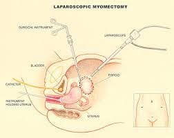
Laparoscopic Myomect
Upto 80% off
90% Rated
Satisfactory

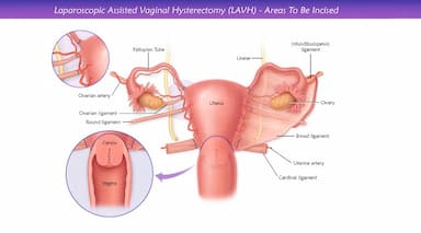
LAVH
Upto 80% off
90% Rated
Satisfactory
NOTE
Upto 80% off
90% Rated
Satisfactory
CABG
Upto 80% off
90% Rated
Satisfactory

Accurate staging of PNETs is essential for determining the best course of treatment and predicting patient outcomes. Staging involves determining the size and location of the tumor, as well as assessing whether the tumor has spread to nearby lymph nodes or other parts of the body. PET scans can be highly effective in staging PNETs because they can detect the presence of metastases in other parts of the body.
One of the most commonly used radioactive tracers for staging PNETs is 68Ga-DOTA-TOC. This tracer binds to somatostatin receptors, which are often overexpressed in PNETs. By binding to these receptors, the tracer can be used to visualize the location and extent of the tumor, as well as detect the presence of metastases.
Several studies have shown that PET scans using 68Ga-DOTA-TOC can be highly effective in staging PNETs. For example, a study published in the Journal of Nuclear Medicine found that 68Ga-DOTA-TOC PET imaging had a sensitivity of 89% and a specificity of 93% in detecting PNET metastases. Another study published in the same style and tone:
The Journal of Clinical Endocrinology and Metabolism found that 68Ga-DOTA-TOC PET imaging had a sensitivity of 93% and a specificity of 95% in detecting PNETs.
In addition to 68Ga-DOTA-TOC, other radioactive tracers have also been used for PET imaging in PNET staging. For example, 18F-fluoro-L-dihydroxyphenylalanine (FDOPA) is a tracer that is taken up by cells that produce dopamine, which is often overexpressed in PNETs. Several studies have shown that FDOPA PET imaging can be highly effective in detecting PNETs and their metastases, with sensitivities ranging from 71% to 100% and specificities ranging from 75% to 100%.
Benefits of PET Scan for PNET Diagnosis and Staging
PET imaging has several advantages over other imaging modalities for PNET diagnosis and staging. One major advantage is its ability to detect metabolic activity in the body, which can be highly informative in detecting and staging PNETs. Additionally, PET imaging is noninvasive and does not require the use of ionizing radiation, making it a safer option than other imaging modalities such as computed tomography (CT) or magnetic resonance imaging (MRI).
Another advantage of PET imaging is its ability to provide whole-body imaging, allowing for the detection of metastases in other parts of the body. This is particularly important for PNETs, which have a high propensity for metastasis to the liver and other organs.
Limitations of PET Scan for PNET Diagnosis and Staging
Despite its many advantages, PET imaging also has several limitations for PNET diagnosis and staging. One major limitation is its relatively low spatial resolution compared to other imaging modalities such as CT or MRI. This can make it difficult to precisely locate small tumors or lesions, which can impact accurate staging and treatment planning.
Another limitation of PET imaging is its inability to distinguish between different types of PNETs. For example, functional PNETs, which produce hormones that can cause specific symptoms, may have different metabolic profiles than non-functional PNETs, which do not produce hormones. This can make it difficult to accurately diagnose and stage different types of PNETs using PET imaging alone.
Conclusion
PET imaging has emerged as a highly effective imaging modality for the diagnosis and staging of PNETs. The ability of PET imaging to detect metabolic activity in the body makes it a valuable tool for detecting and staging PNETs, particularly in cases where other imaging modalities may be inconclusive. While PET imaging has several limitations, its many advantages make it a valuable tool in the fight against PNETs. With continued research and development, PET imaging is likely to become an even more important tool in the diagnosis and treatment of PNETs in the years to come.
Wellness Treatment
Give yourself the time to relax
Lowest Prices Guaranteed!

Lowest Prices Guaranteed!