PET Scan for Gallbladder Cancer: Diagnosis and Staging
17 May, 2023
 Obaidullah Junaid
Gallbladder cancer is a relatively rare but highly aggressive form of cancer that is notoriously difficult to detect and treat. However, with advancements in medical imaging, such as the use of PET scans, the diagnosis, and staging of gallbladder cancer have become more accurate and effective.
PET stands for Positron Emission Tomography, which is a non-invasive imaging technique that uses a radioactive tracer to produce detailed images of the body's internal organs and tissues. PET scans are commonly used to detect and stage various forms of cancer, including gallbladder cancer.
Transform Your Beauty, Boost Your Confidence
Find the right cosmetic procedure for your needs.
We specialize in a wide range of cosmetic procedures

In this blog, we will discuss how PET scans are used in the diagnosis and staging of gallbladder cancer, including their benefits, risks, and limitations.
Diagnosis of Gallbladder Cancer with PET Scans
The diagnosis of gallbladder cancer often begins with a physical exam and medical history review, followed by various imaging tests to determine the location, size, and extent of the tumor. These imaging tests may include ultrasound, CT scans, MRI scans, and PET scans.
PET scans are particularly useful in the diagnosis of gallbladder cancer because they can detect changes in the metabolism of cancerous cells. Cancer cells tend to have a higher metabolic rate than healthy cells, and PET scans can detect this by using a radioactive tracer that is absorbed by the cells and emits gamma rays.
The most commonly used radioactive tracer for PET scans is FDG (fluorodeoxyglucose), which is a type of sugar that is absorbed by cells that require energy, such as cancer cells. Once FDG is absorbed by the cells, it emits gamma rays that can be detected by a PET scanner and used to create a detailed image of the tumor.
During a PET scan for gallbladder cancer, the patient will be injected with a small amount of FDG and then asked to rest for about an hour to allow the tracer to be absorbed by the cells. The patient will then lie on a table and be moved through a PET scanner, which will produce detailed images of the gallbladder and surrounding tissues.
Most popular procedures in
Laparoscopic Cystect
Upto 80% off
90% Rated
Satisfactory
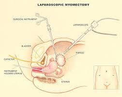
Laparoscopic Myomect
Upto 80% off
90% Rated
Satisfactory
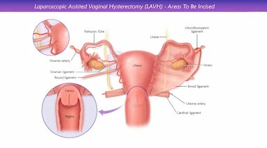
LAVH
Upto 80% off
90% Rated
Satisfactory
NOTE
Upto 80% off
90% Rated
Satisfactory
CABG
Upto 80% off
90% Rated
Satisfactory

The images produced by a PET scan can help doctors determine whether a tumor is cancerous or benign, as well as the size and location of the tumor. PET scans can also help identify whether the cancer has spread to other areas of the body, such as the lymph nodes or liver.
Staging Gallbladder Cancer with PET Scans
Staging refers to the process of determining the extent of a cancerous tumor and whether it has spread to other parts of the body. Staging is important in determining the appropriate treatment options and predicting the patient's prognosis.
PET scans are particularly useful in staging gallbladder cancer because they can detect even small tumors and identify areas of the body where the cancer has spread. PET scans can also provide information about the metabolic activity of the cancer cells, which can help doctors predict the tumor's aggressiveness and potential response to treatment.
In addition to a PET scan, other imaging tests, such as CT scans and MRI scans, may be used to stage gallbladder cancer. These tests can provide detailed images of the tumor and surrounding tissues, as well as identify any areas of the body where the cancer has spread.
Benefits of PET Scans for Gallbladder Cancer
PET scans use a special type of imaging technology that allows doctors to see metabolic activity in the body. Cancerous cells often have different metabolic activity than healthy cells, so PET scans can be used to detect changes in the metabolism of cancerous cells. This makes PET scans a useful tool in the diagnosis and staging of gallbladder cancer.
- Diagnosis of Gallbladder Cancer: PET scans can be used to help diagnose gallbladder cancer by detecting changes in the metabolism of cancerous cells. If a PET scan detects an area of increased metabolic activity in the gallbladder, it may indicate the presence of cancer. A biopsy may be needed to confirm the diagnosis, but a PET scan can provide important information about the location and size of the tumor.
- Staging of Gallbladder Cancer: PET scans can also be used to stage gallbladder cancer by detecting whether the cancer has spread to other parts of the body. PET scans can detect changes in the metabolism of cancerous cells in the liver, lymph nodes, and other areas where gallbladder cancer is likely to spread. This information can help doctors determine the best course of treatment and predict the patient's prognosis.
- Monitoring the Effectiveness of Treatment: PET scans can be used to monitor the effectiveness of treatment for gallbladder cancer. By detecting changes in the metabolism of cancerous cells over time, PET scans can help doctors determine whether the treatment is working and whether the cancer is responding to the therapy. This can help doctors adjust the treatment plan as needed and provide a more accurate prognosis for the patient.
- Minimally Invasive: PET scans are a non-invasive imaging technique, meaning they do not require any incisions or surgical procedures. This makes PET scans a less invasive option for diagnosing and staging gallbladder cancer than other imaging tests, such as CT scans or MRI scans.
Risks of PET Scans for Gallbladder Cancer
While PET scans can be useful in the diagnosis and staging of gallbladder cancer, they also come with some risks that should be considered.
- Exposure to Radiation: PET scans use a small amount of radioactive material to produce images of the body. While the amount of radiation used in a PET scan is considered safe for most people, it can increase the risk of cancer over time. The risk of radiation exposure is higher in children and young adults, and in people who have had multiple imaging tests that use radiation.
- Allergic Reactions: The radioactive tracer used in PET scans can cause an allergic reaction in some people. Symptoms of an allergic reaction may include itching, hives, swelling, and difficulty breathing. Allergic reactions to the radioactive tracer used in PET scans are rare, but it is important to tell your doctor if you have a history of allergies.
- False-Positive or False-Negative Results: PET scans can produce false-positive or false-negative results, which can lead to unnecessary tests or delayed treatment. False-positive results occur when a PET scan indicates the presence of cancer when there is none, while false-negative results occur when a PET scan fails to detect cancer that is present. It is important to discuss the accuracy of PET scans with your doctor and to consider additional tests, such as a biopsy, to confirm a cancer diagnosis.
- Cost: PET scans can be expensive, and not all insurance plans cover the cost of the procedure. The cost of a PET scan can vary depending on the location and type of facility, as well as the specific imaging equipment used. Patients should check with their insurance provider to determine coverage and out-of-pocket costs.
Limitations of PET Scans for Gallbladder Cancer
While PET scans are a valuable tool in the diagnosis and staging of gallbladder cancer, they do have some limitations that should be considered.
- PET scans cannot distinguish between cancerous and non-cancerous lesions. Therefore, additional tests may be needed to confirm a diagnosis.
- PET scans may produce false-positive or false-negative results. False-positive results occur when the PET scan detects a suspicious area that is not cancerous, leading to unnecessary tests or treatment. False-negative results occur when the PET scan does not detect cancerous cells, leading to delayed diagnosis and treatment.
- PET scans are expensive and may not be covered by all insurance plans.
- PET scans are not suitable for all patients, particularly those with certain medical conditions or allergies.
How can we help with the treatment?
If you're on the lookout for treatment in India, Thailand, Singapore, Malaysia, UAE, and Turkey, let Healthtrip be your compass. We will serve as your guide throughout your medical treatment. We'll be by your side, in person, even before your medical journey commences. The following will be provided to you:
- Connect with renowned doctors from a network spanning 35+ countries and access the world's largest health travel platform.
- Collaboration with 335+ top hospitals , including Fortis and Medanta.
- Comprehensive treatments from Neuro to Cardiac to Transplants, Aesthetics, and Wellness.
- Post-treatment care and assistance.
- Teleconsultations at $1/minute with leading surgeons.
- Trusted by 44,000+ patients for appointments, travel, visa, and forex assistance.
- Access top treatments and packages, such as Angiograms and many more.
- Gain insights from genuine patient experiences and testimonials.
- Stay updated with our medical blog.
Our success stories
Conclusion
In summary, PET scans are a valuable tool in the diagnosis and staging of gallbladder cancer. They can detect even small tumors and identify areas of the body where the cancer has spread. PET scans can also provide information about the metabolic activity of the cancer cells, which can help doctors predict the tumor's aggressiveness and potential response to treatment.
While PET scans do have some limitations and risks, they are generally considered safe and effective for patients with suspected gallbladder cancer. Patients should discuss the benefits, risks, and limitations of PET scans with their doctors before undergoing the procedure.
Wellness Treatment
Give yourself the time to relax
Lowest Prices Guaranteed!

Lowest Prices Guaranteed!