PET Scan for Esophageal Cancer: Diagnosis and Staging
13 May, 2023
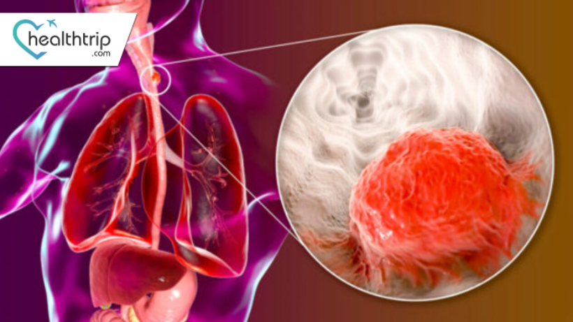
Esophageal cancer is one of the most aggressive cancers and its diagnosis and staging are essential for proper treatment. PET scan is a non-invasive imaging technique that is used for diagnosis, staging, and monitoring of esophageal cancer. PET scan provides detailed information about the size, location, and extent of cancer in the body, which helps doctors in making a treatment plan for the patient. In this blog, we will discuss the PET scan for esophageal cancer in detail.
Understanding Esophageal Cancer
Transform Your Beauty, Boost Your Confidence
Find the right cosmetic procedure for your needs.
We specialize in a wide range of cosmetic procedures

Esophageal cancer is a type of cancer that affects the esophagus, which is the tube that connects the throat to the stomach. The main types of esophageal cancer are squamous cell carcinoma and adenocarcinoma. Esophageal cancer is often diagnosed at an advanced stage when it has spread to other parts of the body. Therefore, early detection and staging are essential for effective treatment.
What is a PET scan?
Positron Emission Tomography (PET) scan is a medical imaging technique that uses a special dye containing radioactive tracers to create images of the inside of the body. PET scan can detect changes in cellular activity and metabolism, which can indicate the presence of disease, including cancer. PET scan is a non-invasive procedure that does not involve any incisions or surgery.
PET Scan for Esophageal Cancer
PET stands for positron emission tomography, which is an imaging technique that uses a small amount of radioactive material to create detailed images of the body. PET scan is often used in combination with other imaging techniques such as CT scan, MRI, and ultrasound.
How PET Scan Works?
Most popular procedures in
Laparoscopic Cystect
Upto 80% off
90% Rated
Satisfactory
Laparoscopic Myomect
Upto 80% off
90% Rated
Satisfactory
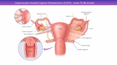
LAVH
Upto 80% off
90% Rated
Satisfactory
NOTE
Upto 80% off
90% Rated
Satisfactory
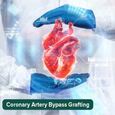
CABG
Upto 80% off
90% Rated
Satisfactory
In PET scan, a small amount of a radioactive substance called a tracer is injected into the patient's body. The tracer is usually a form of sugar, such as glucose, which is taken up by cancer cells faster than normal cells. The tracer emits positrons, which interact with electrons in the body, resulting in the emission of gamma rays. These gamma rays are detected by a PET scanner, which creates a three-dimensional image of the body.
PET Scan Procedure
Before the PET scan, the patient is advised to avoid eating for a few hours. The patient is then injected with the tracer and asked to rest for about an hour to allow the tracer to spread throughout the body. The patient is then placed on a PET scanner, which takes about 30 minutes to create a detailed image of the body.
Benefits of PET Scan for Esophageal Cancer
PET scan has several benefits for esophageal cancer diagnosis and staging.
- PET scan provides detailed information about the location, size, and extent of cancer in the body, which helps doctors in making a treatment plan.
- PET scan is a non-invasive imaging technique, which means that it does not require any incisions or surgery.
- PET scan is a relatively quick procedure and the patient can return to normal activities immediately after the scan.
Limitations of PET Scan for Esophageal Cancer
PET scan has some limitations that should be considered.
- PET scan cannot differentiate between cancer and inflammation, infection, or scar tissue.
- PET scan may not detect small tumours or tumours with low metabolic activity.
- PET scan may produce false-positive or false-negative results, which may require further testing.
PET Scan for Esophageal Cancer Staging
Esophageal cancer staging is the process of determining the extent of cancer in the body. Staging helps doctors in making a treatment plan for the patient. PET scan is often used for esophageal cancer staging, along with other imaging techniques such as CT scan and endoscopic ultrasound.
PET scan Procedure
Before the PET scan procedure, the patient is injected with a small amount of radioactive tracer, which is absorbed by the body's cells. The tracer is typically a form of glucose that is absorbed by cancer cells more rapidly than normal cells, making it easier to detect. The patient is then placed on a table and moved through a large circular scanner, which detects the radioactive tracer and creates images of the inside of the body.
The PET scan procedure is relatively quick and typically takes about 30 minutes to complete. The patient may be asked to avoid eating or drinking for several hours before the procedure to ensure accurate results.
Stages of Esophageal Cancer
Esophageal cancer is staged using the TNM system, which stands for tumour, lymph nodes, and metastasis. The stages of esophageal cancer are:
- Stage 0: Cancer cells are only in the inner layer of the esophagus.
- Stage I: Cancer cells have spread beyond the inner layer of the esophagus but have not spread to nearby lymph nodes or other organs.
- Stage II: Cancer cells have spread to nearby lymph nodes or tissues, but not to distant organs.
- Stage III: Cancer cells have spread to nearby organs and lymph nodes.
- Stage IV: Cancer cells have spread to distant organs such as the liver, lungs, or bones.
PET Scan for Esophageal Cancer Staging
PET scan is used for esophageal cancer staging to determine the extent of cancer in the body. PET scan can detect cancer cells in the lymph nodes and other organs, which helps in determining the stage of cancer. PET scan is often used in combination with other imaging techniques such as CT scan and endoscopic ultrasound for accurate staging of esophageal cancer.
Conclusion
PET scan is a non-invasive imaging technique that is used for the diagnosis and staging of esophageal cancer. This procedure provides detailed information about the location, size, and extent of cancer in the body, which helps doctors in making a treatment plan for the patient. However, PET scan has limitations, such as false-positive or false-negative results, which can lead to inaccurate diagnosis and staging of esophageal cancer.
To address these limitations, PET scan is often used in combination with other imaging techniques, such as CT scan or MRI. CT scan provides detailed information about the anatomy of the esophagus and surrounding tissues, while PET scan provides information about the metabolic activity of the tissues. This combination of imaging techniques can help doctors in accurately diagnosing and staging esophageal cancer.
In summary, PET scan is a valuable tool for the diagnosis and staging of esophageal cancer, but it has limitations that should be considered. Using PET scan in combination with other imaging techniques can increase the accuracy of the diagnosis and staging of esophageal cancer, leading to better treatment outcomes.
Wellness Treatment
Give yourself the time to relax
Lowest Prices Guaranteed!

Lowest Prices Guaranteed!