Read Blogs on Healthtrip
10 Natural Home Remedies for Headache Relief
Headaches are a common ailment that many people experience on a regular basis. They can be caused by a variety of factors, such as tension, dehydration, sinus problems, and even certain foods. While over-the-counter pain relievers can provide temporary relief, they may not always be the best solution. Fortunately, there are several natural home remedies that can help alleviate headache pain.Stay hydrated: Dehydration is a common cause of headaches. Drinking plenty of water throughout the day can help prevent headaches from occurring. Aim for at least eight glasses of water per day, and more if you are physically active or in a hot climate. You can also try drinking other fluids, such as herbal tea or coconut water, to help keep you hydrated.Apply heat or cold: Applying heat or cold to the affected area can help relieve headache pain. You can use a hot or cold compress, depending on your preference. A warm compress can help relax tense muscles and increase blood flow, while a cold compress can help reduce inflammation and numb the area. You can use a heating pad, a warm towel, or a cold pack to apply heat or cold to the affected area.Practice relaxation techniques: Stress and tension can contribute to headaches. Practicing relaxation techniques, such as deep breathing, meditation, or yoga, can help reduce stress and tension and prevent headaches from occurring. You can also try progressive muscle relaxation, which involves tensing and relaxing different muscle groups to promote relaxation.Massage: Massaging the affected area can help relieve headache pain. You can use your fingers to gently massage your temples, forehead, and neck. You can also use a handheld massager or seek out a professional massage therapist for a more thorough massage.Ginger: Ginger is a natural anti-inflammatory that can help reduce headache pain. You can brew ginger tea by steeping grated ginger in boiling water for 10-15 minutes. You can also add ginger to your meals or take ginger supplements.Lavender oil: Lavender oil has calming properties that can help relieve headache pain. You can apply lavender oil to your temples and forehead, or add a few drops to a warm bath to help promote relaxation.Peppermint oil: Peppermint oil has a cooling effect that can help relieve headache pain. You can apply peppermint oil to your temples and forehead, or add a few drops to a diffuser to inhale the scent.Magnesium: Magnesium is an important mineral that can help prevent headaches. You can increase your magnesium intake by eating foods such as almonds, spinach, and avocados, or by taking magnesium supplements.Chamomile tea: Chamomile tea has calming properties that can help relieve headache pain. You can brew chamomile tea by steeping chamomile flowers in boiling water for 10-15 minutes.Exercise: Exercise can help reduce stress and tension, which can help prevent headaches from occurring. You can engage in low-impact exercise such as walking, swimming, or cycling, or try yoga or Pilates. In conclusion, headaches can be a painful and debilitating condition, but there are several natural home remedies that can help alleviate the pain. By staying hydrated, applying heat or cold, practicing relaxation techniques, massaging the affected area, and incorporating foods and supplements such as ginger and magnesium into your diet, you can help prevent headaches from occurring and find relief when they do.It's important to note that while natural home remedies can be effective for some people, they may not work for everyone. If you experience frequent or severe headaches, it's important to talk to your healthcare provider to rule out any underlying medical conditions or to discuss other treatment options.Additionally, some natural remedies may not be suitable for everyone. For example, if you have sensitive skin, using essential oils topically may cause irritation. Always do a patch test before using any new remedy, and consult with your healthcare provider if you have any concerns or questions.In summary, there are several natural home remedies for headache relief, including staying hydrated, applying heat or cold, practicing relaxation techniques, massaging the affected area, incorporating foods and supplements such as ginger and magnesium into your diet, and exercising. It's important to remember that what works for one person may not work for another, so it's important to find what works best for you. If you experience frequent or severe headaches, be sure to talk to your healthcare provider to rule out any underlying medical conditions or to discuss other treatment options.
PET Scan for Uterine Cancer: Diagnosis and Staging
Uterine cancer, also known as endometrial cancer, is the most common cancer of the female reproductive system. It occurs when abnormal cells in the endometrium begin to grow out of control. Early detection and accurate staging of uterine cancer are essential for successful treatment and improved outcomes. PET scans are important tools in the diagnosis and classification of cervical cancer, providing valuable information about the extent and spread of disease.What is a PET scan?PET (Positron Emission Tomography) is a non-invasive imaging technique that uses radioactive tracers to create detailed pictures of organs and tissues inside the body. Tracers are substances that are absorbed by the cells of the body and emit positrons, which are captured by special cameras. The resulting images provide information about the metabolic activity of cells that can be used for cancer diagnosis and classification.How do PET scans work in diagnosing and staging cervical cancer?Diagnosis and staging of uterine cancer usually involves a PET scan after a biopsy confirms the presence of cancer. Before the scan, the patient is injected with a small amount of a radioactive tracer that is taken up by cancer cells in the uterus. The positrons emitted by the tracer are captured by a PET scanner and used to create detailed images of the cancer and surrounding tissue.PET scans can detect the presence of cancer cells in the uterus and surrounding tissues, and can detect cancer that has spread to nearby lymph nodes or other organs. This information is important for determining the stage of cancer and planning effective treatment.Benefits of PET Scans for Diagnosing and Staging Uterine CancerPET scans have the following advantages for diagnosing and staging uterine cancer:1. Improved accuracy: PET scans are more accurate than other imaging tests, such as CT and MRI scans, for detecting the spread of uterine cancer.2. Early detection: A PET scan can detect small tumors and cancer cells in the uterus before they are visible on other imaging tests.3. Improved staging: A PET scan can help determine the stage of cancer by detecting if it has spread to nearby lymph nodes or other organs. 4. Treatment plan: PET scans provide valuable information about the extent and location of the cancer, helping doctors make effective treatment plans.5. Monitoring treatment response: PET scans can be used to monitor the response of uterine cancer to treatment over time.Preparing for a PET scan for cervical cancerPreparing for a PET scan for uterine cancer usually requires following your healthcare provider's instructions. These instructions may include:1. Fasting: Patients may need to fast for several hours before the scan2. Drugs: Patients may need to stop taking certain medications that may affect scan results.3. Relax: Patients may be asked to rest prior to scanning to reduce the risk of motion artefacts in the image.4. Dress: Patients should wear comfortable, loose-fitting clothing for the scan.5. Hydration: Patients should drink plenty of water before and after the scan to flush the tracer out of the body. PET Scan Risks for Uterine CancerPET scans are generally considered safe, but involve low levels of radiation exposure. However, radiation exposure is typically lower than CT scans and his X-rays. Additionally, some people may be allergic to tracers used in PET scans. Symptoms of an allergic reaction may include hives, itching, or difficulty breathing.ConclusionPET scanning is an important tool for diagnosing and staging uterine cancer. Improved accuracy, early detection, improved staging, treatment planning, Monitoring response to treatment. PET scans are especially useful for detecting small tumours and cancer cells in the uterus before they can be seen by other imaging tests. It also helps determine the extent of the cancer and whether it has spread to nearby lymph nodes or other organs.If you've been diagnosed with uterine cancer, talk to your doctor about whether a PET scan is right for your situation. PET scans provide valuable information about the extent and spread of cancer, helping guide treatment decisions and improve outcomes. With early detection and accurate staging, uterine cancer can be successfully treated and many women live longer, healthier lives.
PET Scan for Vulvar Cancer: Diagnosis and Staging
Vulvar cancer is a rare but serious type of cancer that affects the external female genitalia, specifically the vulva. It is estimated that approximately 6,000 women in the United States are diagnosed with vulvar cancer each year. Like other types of cancer, early detection and accurate staging are crucial for determining the most effective treatment plan and improving the patient's chances of survival. PET scans are a valuable tool in the diagnosis and staging of vulvar cancer.What is a PET scan for vulvar cancer?A PET scan for vulvar cancer is an imaging test that uses a radioactive tracer to visualise areas of the body with abnormal metabolic activity. The tracer is injected into the patient's bloodstream, where it is absorbed by tissues that are actively using glucose, such as cancer cells. The PET scan then creates images of these areas of metabolic activity, allowing doctors to identify and stage vulvar cancer.How is a PET scan used to diagnose and stage vulvar cancer?PET scans are often used in conjunction with other imaging tests, such as CT scans or MRI scans, to diagnose and stage vulvar cancer. By analysing the images produced by these tests, doctors can determine the size and location of the tumour, as well as whether it has spread to other parts of the body, such as nearby lymph nodes or distant organs. This information is crucial for determining the most effective treatment plan for the patient.Are there any risks or side effects associated with PET scans for vulvar cancer?Like any medical procedure, PET scans for vulvar cancer do carry some risks and potential side effects, such as radiation exposure, allergic reactions, and false positive or false negative results. However, these risks are generally considered to be minimal, and the benefits of using PET scans to diagnose and stage vulvar cancer typically outweigh the risks.How do I prepare for a PET scan for vulvar cancer?Patients may be instructed to avoid eating or drinking for a certain period of time prior to the PET scan, as well as to avoid certain medications that may interfere with the test results. Patients should also inform their doctor if they are pregnant or breastfeeding, as the radioactive tracer used in the PET scan can potentially harm a developing foetus or be passed through breast milk.What happens during a PET scan for vulvar cancer?During a PET scan for vulvar cancer, the patient will receive an injection of a small amount of radioactive tracer. The tracer will then be absorbed by tissues in the body that are actively using glucose, such as cancer cells. After a short waiting period, the patient will lie down on a table and be moved into a scanner, which will create images of the areas of metabolic activity in the body.How long does a PET scan for vulvar cancer take?The actual scan typically takes between 30 and 60 minutes, although the entire process may take several hours from start to finish. Patients may also be required to stay at the medical facility for a short period of time following the scan to ensure that there are no adverse reactions to the tracer.How soon will I receive the results of my PET scan for vulvar cancer?The timing of the results of a PET scan for vulvar cancer can vary depending on the medical facility and the specific circumstances of the patient's case. In some cases, the results may be available within a few days, while in other cases, it may take several weeks for the results to be fully analysed and reported to the patient.What are the benefits of PET scans for vulvar cancer?PET scans are a valuable tool in the diagnosis and staging of vulvar cancer, as they can provide detailed information about the location and extent of the cancer. This information is crucial for determining the most effective treatment plan for the patient, which may include surgery, chemotherapy, radiation therapy, or a combination of these approaches.In addition, PET scans can also help monitor the effectiveness of treatment over time. By performing follow-up PET scans, doctors can determine whether the cancer has responded to treatment, and make adjustments to the treatment plan if necessary.Are there any limitations to PET scans for vulvar cancer?While PET scans are a valuable tool in the diagnosis and staging of vulvar cancer, they do have some limitations. For example, PET scans are not always able to distinguish between cancerous and non-cancerous tissue, which can sometimes lead to false positive results. Additionally, PET scans may not be able to detect very small tumors, or tumors that have not yet begun to exhibit abnormal metabolic activity.Overall, however, PET scans are a valuable diagnostic tool for vulvar cancer, and can provide important information that can help guide treatment decisions and improve patient outcomes. If you have been diagnosed with vulvar cancer or are at high risk for the disease, talk to your doctor about whether a PET scan may be right for you. With early detection and accurate staging, vulvar cancer can be effectively treated, and patients can go on to live healthy, fulfilling lives.
PET scan for Testicular Cancer: Diagnosis and Staging
Testicular cancer is a rare but treatable cancer that affects the testicles, the male reproductive organs responsible for sperm and testosterone production. In recent years, the use of his PET scans in the diagnosis and staging of testicular cancer has become more popular. This article reviews the use of PET scanning in the diagnosis and staging of testicular cancer. What is a PET scan?PET stands for Positron Emission Tomography and is a type of imaging test that uses a small amount of radioactive material to create pictures inside the body. In a PET scan, the patient is injected with a small amount of radioactive material called a tracer. This tracer then travels throughout the body and accumulates in areas of high metabolic activity. B. Cancer cells. A PET scanner then detects the radioactive material and creates detailed pictures of the inside of your body. PET scan to diagnose testicular cancerTesticular cancer is usually diagnosed using a combination of physical examination, blood tests, and imaging tests. A PET scan is one of the imaging tests that can be used to diagnose testicular cancer. PET scans are often used to determine the stage of the cancer and its spread after testicular cancer has already been diagnosed. However, in some cases, a PET scan can also be used as part of the initial diagnostic workup. Injects a patient with a radioactive tracer during his PET scan for testicular cancer. This tracer moves through the body and accumulates in areas of high metabolic activity. These areas include not only cancer cells in the testicles, but also other areas of the body where cancer has spread. PET scanners then create detailed images of the inside of the body, helping doctors identify areas of abnormal activity that may indicate testicular cancer. PET scans are especially useful for detecting cancer that has spread to other parts of the body, such as lymph nodes, lungs, or liver. PET scan for staging testicular cancerOnce testicular cancer has been diagnosed; the next step is to determine the stage of the cancer. Staging is the process of determining how advanced the cancer is and how far it has spread. PET scans are often used to stage testicular cancer because they help identify areas of the body where cancer has spread. This information is important in determining the most appropriate treatment plan for the patient. A PET scan helps doctors determine if cancer has spread to nearby lymph nodes or other organs, such as the lungs or liver. This information is used to assign a stage to the cancer, ranging from stage 1 (early stage) to stage 4 (advanced stage). PET scans can also help monitor the progress of testicular cancer treatment. They help doctors determine whether the cancer has responded to treatment or whether it continues to grow or spread. Benefits of PET Scans for Testicular CancerUsing PET scans for the diagnosis and grading of testicular cancer has several advantages. These benefits are: 1. Accurate diagnosis:A PET scan can help identify areas of abnormal activity that may indicate testicular cancer, even when other imaging tests are inconclusive.2. Staging:A PET scan can help determine the stage of testicular cancer and identify areas where the cancer has spread. This information is important in developing an appropriate treatment plan.3. Treatment monitoring:PET scans help monitor the progress of testicular cancer treatment and determine if the cancer has responded to treatment.4. Minimal Risk:PET scans are generally considered safe and have minimal risks, especially when compared to other imaging modalities that use high levels of radiation. PET risks and side effectsAs with any medical procedure, PET scans come with some risks and potential side effects. However, these risks are generally considered minimal, especially when compared to the potential benefits of using his PET scans in diagnosing and treating testicular cancer.Potential risks and side effects of PET scans include: 1. Radiation exposure:PET scans use a small amount of radioactive material and therefore expose the patient to a small amount of radiation. However, the amount of radiation used in PET scans is generally considered safe, and the risks of radiation exposure usually outweigh the potential benefits of the procedure. 2. Allergic reactions: Some patients may have allergic reactions to the radiotracers used in PET scans. Symptoms of an allergic reaction include itching, hives, difficulty breathing, and swelling of the face, lips, tongue, and throat. However, these reactions are rare. 3. Discomfort or pain: Some patients may experience discomfort or pain at the injection site where the radiotracer was administered. However, this discomfort is usually mild and temporary. 4. False Positives: As with any imaging test, PET scans can give false-positive results. This means that the scan can show areas of abnormal activity that aren't actually cancer. In some cases, this can lead to unnecessary follow-up tests and treatments. 5. False negatives: PET scans can also give false negative results. This means that active areas of cancer may not show up in the scan. This can occur when the cancer is in its early stages or when the cancer is not very metabolically active. It's important to note that the benefits of using his PET scans to diagnose and treat testicular cancer typically outweigh the risks and potential side effects. Patients considering a PET scan should discuss the potential risks and benefits with their physician and make an informed decision whether to have a PET scan. CONCLUSIONPET scanning is a powerful tool in the diagnosis and staging of testicular cancer. These scans help doctors identify areas of abnormal metabolic activity that may indicate cancer and help determine the stage of the cancer and whether it has spread to other parts of the body. PET scans have some risks and potential side effects, but these risks are generally considered minimal. The benefits of using her PET scans to diagnose and treat testicular cancer usually outweigh the risks. Patients considering a PET scan should discuss the potential risks and benefits with their doctor and make an informed decision about whether to have surgery.
PET scan for Renal Cancer: Diagnosis and Staging
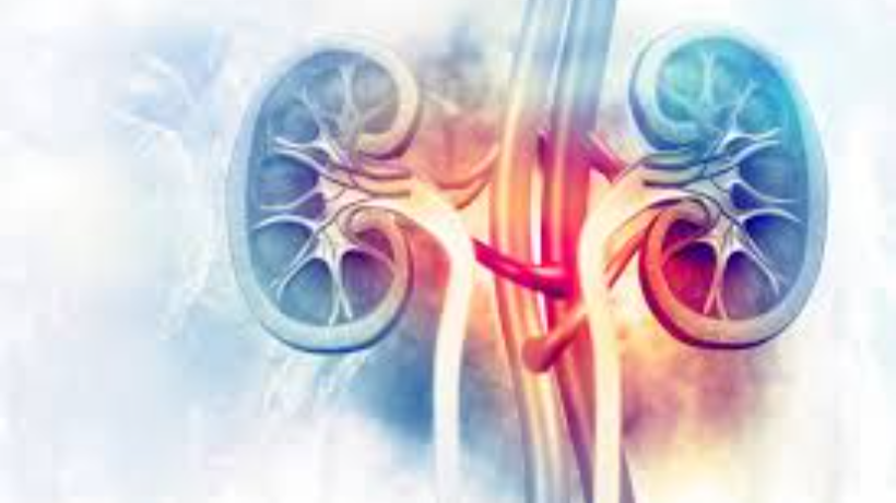
PET scan (Positron Emission Tomography) is a diagnostic imaging technique that uses a radioactive tracer to produce images of the internal organs and tissues of the body. PET scans are widely used in the diagnosis and staging of various types of cancer, including renal (kidney) cancer.Renal cancer is a type of cancer that develops in the cells of the kidney. It is the seventh most common cancer in men and the ninth most common in women worldwide. According to the American Cancer Society, about 76,080 new cases of kidney cancer will be diagnosed in the United States in 2021, and about 13,780 people will die from this disease.Diagnosis of renal cancer typically involves a combination of medical history, physical examination, blood tests, imaging studies, and biopsy. PET scans are increasingly being used to diagnose and stage renal cancer, as they provide detailed information about the metabolic activity of the tumour and surrounding tissues.In this blog, we will discuss how PET scans are used in the diagnosis and staging of renal cancer, their benefits, and potential risks.How Does a PET Scan Work?Before discussing the use of PET scans in renal cancer diagnosis, it is essential to understand how they work. PET scans involve the use of a small amount of radioactive material called a tracer, which is injected into the bloodstream. The tracer travels through the body and accumulates in areas of high metabolic activity, such as tumours.Once the tracer has accumulated in the target tissue, a special camera detects the radiation emitted by the tracer and produces three-dimensional images of the tissue. These images can be used to assess the size, location, and metabolic activity of the tumour, as well as the spread of cancer to other parts of the body.PET scan for Renal Cancer DiagnosisRenal cancer can be challenging to diagnose, as it often has no symptoms in its early stages. When symptoms do occur, they can include blood in the urine, pain or discomfort in the side or lower back, or a lump or mass in the abdomen.To diagnose renal cancer, healthcare providers typically begin with a physical exam and medical history. Blood and urine tests may be used to detect the presence of substances produced by kidney cancer cells. Imaging studies, such as ultrasound, CT scan, or MRI, may be used to visualise the tumour and surrounding tissues.PET scans are increasingly being used to diagnose renal cancer, especially in cases where the tumour is difficult to visualise with other imaging techniques. PET scans can detect the metabolic activity of cancer cells, which may be higher than that of normal cells.A study published in the Journal of Nuclear Medicine found that PET scans are more accurate in detecting renal cancer than CT scans. The study found that PET scans detected 93.1% of renal tumours, compared to 77.8% detected by CT scans.PET scan for Renal Cancer StagingStaging refers to the process of determining the extent and spread of cancer. Accurate staging is essential for determining the most appropriate treatment approach and predicting the prognosis of the disease.Staging of renal cancer typically involves a combination of imaging studies, including CT scans, MRI, and PET scans. PET scans are increasingly being used in renal cancer staging, as they provide detailed information about the metabolic activity of the tumour and surrounding tissues.PET scans can detect the presence of cancer cells in other parts of the body, such as the lymph nodes, bones, or lungs. This is important because renal cancer can spread to other parts of the body, even in its early stages.A study published in the Journal of Urology found that PET scans were highly accurate in detecting the spread of renal cancer to other parts of the body. The study found that PET scans had a sensitivity of 92% and a specificity of 93%, meaning they were highly accurateBenefits of PET Scans for Renal Cancer Diagnosis and StagingPET scans have several benefits for the diagnosis and staging of renal cancer. These include:1. Increased Accuracy: PET scans are more accurate than other imaging techniques in detecting renal cancer, especially in cases where the tumour is difficult to visualise with other imaging techniques.2. Early Detection: PET scans can detect renal cancer in its early stages, before it spreads to other parts of the body.3. Better Staging: PET scans provide detailed information about the metabolic activity of the tumour and surrounding tissues, allowing for more accurate staging of the cancer.4. Treatment Planning: PET scans can help healthcare providers determine the most appropriate treatment approach based on the size, location, and spread of the tumour.5. Monitoring: PET scans can be used to monitor the response of renal cancer to treatment over time. Potential Risks of PET ScansWhile PET scans are generally considered safe, they do involve exposure to a small amount of radiation. However, the amount of radiation exposure is typically less than that of a CT scan or X-ray. Additionally, some people may have an allergic reaction to the tracer used in the PET scan. Symptoms of an allergic reaction can include hives, itching, or difficulty breathing. Preparing for a PET scan for Renal Cancer Preparing for a PET scan for renal cancer typically involvesfollowing some instructions provided by the healthcare provider. These instructions may include:1. Fasting: Depending on the type of tracer used, patients may need to fast for several hours before the scan.2. Medications: Patients may need to stop taking certain medications before the scan, as they may interfere with the results.3. Rest: Patients may be advised to rest before the scan to reduce the risk of motion artefacts in the images.4. Clothing: Patients should wear comfortable, loose-fitting clothing to the scan. Metal objects, such as jewellery or clothing with metal zippers, should be removed.5. Hydration: Patients should drink plenty of water before and after the scan to help flush the tracer out of the body.ConclusionPET scans are increasingly being used in the diagnosis and staging of renal cancer. They provide detailed information about the metabolic activity of the tumour and surrounding tissues, allowing for more accurate diagnosis and staging. PET scans can also help healthcare providers determine the most appropriate treatment approach based on the size, location, and spread of the tumour. While PET scans are generally considered safe, they do involve exposure to a small amount of radiation. Patients should follow the instructions provided by their healthcare provider to prepare for the scan and minimise any potential risks.
PET Scan for Penile Cancer: Diagnosis and Staging
Penile cancer is a rare cancer that affects the penis. Diagnosis is difficult, but early detection is the key to successful treatment. PET scans play an important role in diagnosing and grading penile cancer, allowing healthcare professionals to accurately determine the extent and spread of the cancer and to plan effective treatment.How a PET scan worksPositron Emission Tomography (PET) scans use small amounts of radioactive tracers to make detailed pictures of organs and tissues inside the body. The tracer is usually injected into an arm vein and absorbed by the body's cells, including cancer cells. When the tracer is taken up by the cell, positrons are emitted and captured by the PET scanner. The scanner makes detailed pictures of your body so your health care provider can see the location and extent of cancer cells.PET scans are often used in combination with CT (Computed Tomography) scans, which provide a detailed picture of the internal structures of the body. Two imaging tests are often combined to create a very detailed picture of the body, allowing health care providers to accurately diagnose and classify penile cancer.Diagnosing Penile Cancer with a PET scanOne of the main uses of PET scans for penile cancer is to diagnose cancer and locate it within the penis. PET scans can detect cancer cells in the penis before they are visible with other imaging tests, allowing for early diagnosis and treatment.During a PET scan, a radioactive tracer is absorbed by cancer cells in the penis. The tracer emits positrons, which are captured by a PET scanner and used to create detailed images of cancer cells. This information helps health care providers determine the location and size of the cancer and how far it has spread.Staging Penile Cancer by PET scanOnce penile cancer has been diagnosed, the next step is to determine the stage of the cancer. Staging refers to how far the cancer has spread throughout the body and plays an important role in determining the most effective treatment plan.PET scans are an important tool in staging penile cancer because they can detect cancer that has spread to nearby lymph nodes or other organs. During the scan, the radioactive tracer is absorbed not only by cancer cells in the penis, but also by cancer cells that have spread to other parts of the body.Images produced by PET scans help health care providers determine the stage of cancer and plan effective treatment. For example, if cancer has spread to nearby lymph nodes, surgery may be needed to remove the affected lymph nodes. If cancer has spread to other organs, chemotherapy or radiation therapy may be recommended. Preparing for a PET scan for penile cancerPreparing for a PET scan for penile cancer involves following your healthcare provider's instructions. Patients may need to fast for several hours before the scan, stop taking certain medications, and drink plenty of water to wash the tracer out of the body. Although PET scans are generally considered safe, they involve low levels of radiation exposure and some people may be allergic to the tracers used. Patients should discuss any concerns with their doctor before having a PET scan.Penile cancer is a rare but serious condition that is difficult to diagnose and treat. PET scans play an important role in diagnosing and grading penile cancer, allowing healthcare professionals to accurately determine the extent and spread of the cancer and to plan effective treatment. If you have been diagnosed with penile cancer, talk to your doctor about whether a PET scan is right for your situation. PET scans provide valuable information about the location and extent of cancer, helping guide and improve treatment decisions result. With early diagnosis and treatment, the outlook for penile cancer is generally good, and PET scans can facilitate this process.A PET scan is just one tool in the diagnosis and treatment of penile cancer. Other imaging tests, such as CT scans and MRIs, can also be used to diagnose and classify cancer. In addition, your health care provider may perform a physical examination and biopsy of affected tissue to confirm the diagnosis.It is important for men to be aware of the signs and symptoms of penile cancer.A lump or growth on the penisSkin changes on the penis, such as redness and thickeningPenile pain or discomfortBleeding or discharge from the penisIf you notice any of these symptoms, it's important to see your doctor right away. Early detection and treatment can help improve outcomes and reduce the risk of complications.In conclusion, PET scanning can be a valuable tool in the diagnosis and staging of penile cancer. They help health care providers identify the location and extent of cancer, which can guide treatment decisions and improve outcomes. Talk to your doctor about whether it's right for your situation. With early diagnosis and treatment, the outlook for penile cancer is generally good, and PET scans can facilitate this process.
PET Scan for Bone Cancer: Diagnosis and Staging
Cancer is a disease that affects millions of people around the world. It is a disease that can strike anyone, regardless of age, gender, or lifestyle. One type of cancer that can be particularly challenging to diagnose and treat is bone cancer. However, with advancements in medical technology, PET scans have become an important tool in diagnosing and staging bone cancer.What is Bone Cancer?Bone cancer is a type of cancer that affects the bones. It is a relatively rare form of cancer, accounting for less than 1% of all cancers. Bone cancer can occur in any bone in the body but is most commonly found in the long bones of the arms and legs.There are two main types of bone cancer: primary bone cancer and secondary bone cancer. Primary bone cancer starts in the bone itself, while secondary bone cancer is cancer that has spread from another part of the body to the bone.Symptoms of bone cancer can include pain, swelling, and a lump or mass in the affected area. These symptoms can often be mistaken for other conditions, which can make diagnosing bone cancer difficult.Diagnosing Bone CancerThe first step in diagnosing bone cancer is a physical examination. During the exam, the doctor will look for any lumps or masses in the affected area and may perform imaging tests to get a closer look at the affected bone.Imaging tests that may be used to diagnose bone cancer include X-rays, CT scans, and MRI scans. These tests can provide detailed images of the bone and can help the doctor determine if there is a tumor present.However, even with these imaging tests, it can be difficult to determine if a tumor is cancerous or not. This is where PET scans come in.What is a PET Scan?PET stands for positron emission tomography. A PET scan is a type of imaging test that uses a radioactive substance called a tracer to look for changes in the body at the cellular level.During a PET scan, the patient is injected with a small amount of radioactive tracer. The tracer travels through the body and is absorbed by cells that are actively growing and dividing, such as cancer cells.As the tracer is absorbed by these cells, it emits positrons, which are small particles that can be detected by the PET scanner. The scanner creates a three-dimensional image of the body that shows where the tracer has accumulated.PET Scan for Bone CancerPET scans can be particularly useful in diagnosing bone cancer because they can detect cancer cells that may not be visible on other imaging tests. PET scans can also help determine the extent of the cancer and whether it has spread to other parts of the body.When used to diagnose bone cancer, a PET scan is often combined with a CT scan. This is known as a PET-CT scan. The CT scan provides detailed images of the bones, while the PET scan shows areas of increased metabolic activity, which can indicate the presence of cancer.Staging Bone CancerStaging is the process of determining how advanced a cancer is and how far it has spread. Staging is an important part of the cancer treatment process because it helps doctors determine the best course of treatment.PET scans can be particularly useful in staging bone cancer because they can detect cancer cells that may not be visible on other imaging tests. This can help doctors determine the extent of the cancer and whether it has spread to other parts of the body.The most common system used to stage bone cancer is the TNM system. This system uses three key pieces of information to determine the stage of the cancer:Tumor (T): This refers to the size of the tumor and whether it has grown into nearby tissues.Nodes (N): This refers to whether the cancer has spread to nearby lymph nodes.Metastasis (M): This refers to whether the cancer has spread to other parts of the body.PET scans can provide valuable information on all three of these factors. They can show the size and location of the primary tumor, as well as any lymph nodes that may be affected. PET scans can also detect any areas of increased metabolic activity in other parts of the body, which can indicate the presence of metastases.Using this information, doctors can determine the stage of the cancer and develop an appropriate treatment plan.Benefits of PET Scans for Bone CancerThere are several benefits of using PET scans for diagnosing and staging bone cancer:Early detection: PET scans can detect cancer cells that may not be visible on other imaging tests, allowing for early detection and treatment.Accurate staging: PET scans can provide detailed information on the extent of the cancer and whether it has spread to other parts of the body, allowing for accurate staging.Personalized treatment: By accurately staging the cancer, doctors can develop a personalized treatment plan that is tailored to the individual patient's needs.Monitoring treatment progress: PET scans can be used to monitor the effectiveness of treatment and detect any new areas of cancer growth.Reduced need for invasive procedures: PET scans can reduce the need for invasive procedures, such as biopsies, by providing detailed information on the location and extent of the cancer.Limitations of PET Scans for Bone CancerWhile PET scans are a valuable tool in diagnosing and staging bone cancer, they do have some limitations:False positives: PET scans can sometimes detect areas of increased metabolic activity that are not cancerous, leading to false positives.False negatives: PET scans can also miss small areas of cancer growth, leading to false negatives.Radiation exposure: PET scans involve exposure to a small amount of radiation, which can increase the risk of cancer over time.Cost: PET scans can be expensive, and not all insurance plans cover the cost.ConclusionPET scans are a valuable tool in diagnosing and staging bone cancer. They can provide detailed information on the location and extent of the cancer, allowing doctors to develop personalized treatment plans that are tailored to the individual patient's needs. While PET scans do have some limitations, their benefits outweigh their risks in most cases. If you or a loved one has been diagnosed with bone cancer, talk to your doctor about whether a PET scan is right for you.
PET Scan for Gallbladder Cancer: Diagnosis and Staging
Gallbladder cancer is a relatively rare but highly aggressive form of cancer that is notoriously difficult to detect and treat. However, with advancements in medical imaging, such as the use of PET scans, the diagnosis, and staging of gallbladder cancer have become more accurate and effective.PET stands for Positron Emission Tomography, which is a non-invasive imaging technique that uses a radioactive tracer to produce detailed images of the body's internal organs and tissues. PET scans are commonly used to detect and stage various forms of cancer, including gallbladder cancer.In this blog, we will discuss how PET scans are used in the diagnosis and staging of gallbladder cancer, including their benefits, risks, and limitations.Diagnosis of Gallbladder Cancer with PET ScansThe diagnosis of gallbladder cancer often begins with a physical exam and medical history review, followed by various imaging tests to determine the location, size, and extent of the tumor. These imaging tests may include ultrasound, CT scans, MRI scans, and PET scans.PET scans are particularly useful in the diagnosis of gallbladder cancer because they can detect changes in the metabolism of cancerous cells. Cancer cells tend to have a higher metabolic rate than healthy cells, and PET scans can detect this by using a radioactive tracer that is absorbed by the cells and emits gamma rays.The most commonly used radioactive tracer for PET scans is FDG (fluorodeoxyglucose), which is a type of sugar that is absorbed by cells that require energy, such as cancer cells. Once FDG is absorbed by the cells, it emits gamma rays that can be detected by a PET scanner and used to create a detailed image of the tumor.During a PET scan for gallbladder cancer, the patient will be injected with a small amount of FDG and then asked to rest for about an hour to allow the tracer to be absorbed by the cells. The patient will then lie on a table and be moved through a PET scanner, which will produce detailed images of the gallbladder and surrounding tissues.The images produced by a PET scan can help doctors determine whether a tumor is cancerous or benign, as well as the size and location of the tumor. PET scans can also help identify whether the cancer has spread to other areas of the body, such as the lymph nodes or liver.Staging Gallbladder Cancer with PET ScansStaging refers to the process of determining the extent of a cancerous tumor and whether it has spread to other parts of the body. Staging is important in determining the appropriate treatment options and predicting the patient's prognosis.PET scans are particularly useful in staging gallbladder cancer because they can detect even small tumors and identify areas of the body where the cancer has spread. PET scans can also provide information about the metabolic activity of the cancer cells, which can help doctors predict the tumor's aggressiveness and potential response to treatment.In addition to a PET scan, other imaging tests, such as CT scans and MRI scans, may be used to stage gallbladder cancer. These tests can provide detailed images of the tumor and surrounding tissues, as well as identify any areas of the body where the cancer has spread.Benefits of PET Scans for Gallbladder CancerPET scans use a special type of imaging technology that allows doctors to see metabolic activity in the body. Cancerous cells often have different metabolic activity than healthy cells, so PET scans can be used to detect changes in the metabolism of cancerous cells. This makes PET scans a useful tool in the diagnosis and staging of gallbladder cancer.Diagnosis of Gallbladder Cancer: PET scans can be used to help diagnose gallbladder cancer by detecting changes in the metabolism of cancerous cells. If a PET scan detects an area of increased metabolic activity in the gallbladder, it may indicate the presence of cancer. A biopsy may be needed to confirm the diagnosis, but a PET scan can provide important information about the location and size of the tumor.Staging of Gallbladder Cancer: PET scans can also be used to stage gallbladder cancer by detecting whether the cancer has spread to other parts of the body. PET scans can detect changes in the metabolism of cancerous cells in the liver, lymph nodes, and other areas where gallbladder cancer is likely to spread. This information can help doctors determine the best course of treatment and predict the patient's prognosis.Monitoring the Effectiveness of Treatment: PET scans can be used to monitor the effectiveness of treatment for gallbladder cancer. By detecting changes in the metabolism of cancerous cells over time, PET scans can help doctors determine whether the treatment is working and whether the cancer is responding to the therapy. This can help doctors adjust the treatment plan as needed and provide a more accurate prognosis for the patient.Minimally Invasive: PET scans are a non-invasive imaging technique, meaning they do not require any incisions or surgical procedures. This makes PET scans a less invasive option for diagnosing and staging gallbladder cancer than other imaging tests, such as CT scans or MRI scans.Risks of PET Scans for Gallbladder CancerWhile PET scans can be useful in the diagnosis and staging of gallbladder cancer, they also come with some risks that should be considered.Exposure to Radiation: PET scans use a small amount of radioactive material to produce images of the body. While the amount of radiation used in a PET scan is considered safe for most people, it can increase the risk of cancer over time. The risk of radiation exposure is higher in children and young adults, and in people who have had multiple imaging tests that use radiation.Allergic Reactions: The radioactive tracer used in PET scans can cause an allergic reaction in some people. Symptoms of an allergic reaction may include itching, hives, swelling, and difficulty breathing. Allergic reactions to the radioactive tracer used in PET scans are rare, but it is important to tell your doctor if you have a history of allergies.False-Positive or False-Negative Results: PET scans can produce false-positive or false-negative results, which can lead to unnecessary tests or delayed treatment. False-positive results occur when a PET scan indicates the presence of cancer when there is none, while false-negative results occur when a PET scan fails to detect cancer that is present. It is important to discuss the accuracy of PET scans with your doctor and to consider additional tests, such as a biopsy, to confirm a cancer diagnosis.Cost: PET scans can be expensive, and not all insurance plans cover the cost of the procedure. The cost of a PET scan can vary depending on the location and type of facility, as well as the specific imaging equipment used. Patients should check with their insurance provider to determine coverage and out-of-pocket costs.Limitations of PET Scans for Gallbladder CancerWhile PET scans are a valuable tool in the diagnosis and staging of gallbladder cancer, they do have some limitations that should be considered.PET scans cannot distinguish between cancerous and non-cancerous lesions. Therefore, additional tests may be needed to confirm a diagnosis.PET scans may produce false-positive or false-negative results. False-positive results occur when the PET scan detects a suspicious area that is not cancerous, leading to unnecessary tests or treatment. False-negative results occur when the PET scan does not detect cancerous cells, leading to delayed diagnosis and treatment.PET scans are expensive and may not be covered by all insurance plans.PET scans are not suitable for all patients, particularly those with certain medical conditions or allergies.ConclusionIn summary, PET scans are a valuable tool in the diagnosis and staging of gallbladder cancer. They can detect even small tumors and identify areas of the body where the cancer has spread. PET scans can also provide information about the metabolic activity of the cancer cells, which can help doctors predict the tumor's aggressiveness and potential response to treatment.While PET scans do have some limitations and risks, they are generally considered safe and effective for patients with suspected gallbladder cancer. Patients should discuss the benefits, risks, and limitations of PET scans with their doctors before undergoing the procedure.
PET Scan for Adrenal Tumor: Diagnosis and Staging
Adrenal tumors are growths that develop in the adrenal glands. These glands are located on top of each kidney and are responsible for producing hormones that regulate a number of bodily functions, including blood pressure, metabolism, and the body's response to stress. While most adrenal tumors are benign, some can be cancerous and require prompt diagnosis and treatment.A PET (positron emission tomography) scan is a powerful imaging tool that can be used to detect and stage adrenal tumors. In this article, we'll take a closer look at how PET scans work and how they can be used in the diagnosis and staging of adrenal tumors.How PET Scans WorkA PET scan uses a small amount of a radioactive substance, called a tracer, to visualize the body's internal organs and tissues. The tracer is injected into the patient's bloodstream and accumulates in areas of the body where there is high metabolic activity. Cancer cells, for example, tend to be more metabolically active than healthy cells and can therefore be detected more easily using PET imaging.Once the tracer has been injected, the patient lies down on a table that slides into a large, donut-shaped machine. This machine contains detectors that can detect the radiation emitted by the tracer. As the patient lies still, the machine rotates around them, collecting data from different angles. This data is then processed by a computer to create detailed, 3D images of the body's internal structures.PET scans can be used to detect a wide range of cancers, including those in the adrenal glands. They can also be used to stage cancers, which means determining the extent to which the cancer has spread beyond its original site.Diagnosing Adrenal Tumors with PET ScansAdrenal tumors can be difficult to diagnose because they often don't cause any symptoms. In some cases, they may be discovered incidentally during an imaging test for another condition. When a patient does present with symptoms, they can include high blood pressure, weight gain, and muscle weakness, among others.If a doctor suspects that a patient may have an adrenal tumor, they may order a PET scan to help confirm the diagnosis. During the scan, the tracer will accumulate in any areas of the body where there is high metabolic activity, including in any tumors that may be present in the adrenal glands.Because PET imaging can detect even small amounts of cancerous tissue, it is highly sensitive and can be very effective in detecting adrenal tumors. In addition, PET scans can be used to differentiate between benign and malignant tumors. Benign tumors tend to have a lower metabolic activity and will therefore show up as less intense on the PET scan than malignant tumors.Staging Adrenal Tumors with PET ScansIn addition to diagnosing adrenal tumors, PET scans can also be used to stage them. Staging refers to the process of determining how far a cancer has spread beyond its original site. This information is critical in determining the most appropriate course of treatment.During a PET scan, the tracer will accumulate not only in the adrenal glands but also in any other areas of the body where there may be cancerous cells. This can include nearby lymph nodes or other organs, such as the liver or lungs.By analyzing the patterns of tracer uptake on the PET scan, doctors can determine the stage of the cancer. For example, if the tracer accumulates only in the adrenal gland, the cancer may be in an early stage and may be treatable with surgery. If the tracer accumulates in nearby lymph nodes or other organs, however, the cancer may be more advanced and may require a more aggressive treatment approach, such as chemotherapy or radiation therapy.Why is a PET scan used for adrenal tumors?Adrenal tumors can be difficult to diagnose and stage using traditional imaging tests such as computed tomography (CT) or magnetic resonance imaging (MRI). These tests can show the size and location of the tumor, but they can't always determine whether the tumor is benign or malignant.PET scans, on the other hand, can provide important information about the metabolism of the tumor cells. Because cancer cells tend to be more active than healthy cells, they absorb more glucose (a type of sugar) from the bloodstream. This increased glucose uptake can be detected by the radiotracer used in the PET scan.By analyzing the distribution of the radiotracer in the body, doctors can determine whether the adrenal tumor is benign or malignant. They can also determine the stage of the tumor, which refers to how advanced it is and whether it has spread to other parts of the body.PET scans can be particularly useful in detecting small tumors that may not be visible on other imaging tests. They can also help doctors monitor the effectiveness of treatment for adrenal tumors by showing changes in the metabolism of the tumor cells over time.What are the risks of a PET scan?PET scans are generally safe, but they do involve a small amount of radiation exposure. The amount of radiation used in a PET scan is similar to that used in other imaging tests such as CT scans, but because PET scans are more sensitive, they may require a higher dose of radiation.The risk of radiation exposure from a PET scan is generally considered to be very low, and the benefits of the test often outweigh the risks. However, women who are pregnant or breastfeeding should avoid PET scans if possible, as radiation can be harmful to developing fetuses and infants.Patients may also be allergic to the radiotracer used in the PET scan, although this is rare. Patients should inform their doctor if they have any allergies or previous adverse reactions to similar tests.In addition, PET scans can produce false positive or false negative results. A false positive result occurs when the scan indicates the presence of cancer when there is none. This can lead to unnecessary tests and procedures. A false negative result occurs when the scan does not detect cancer that is present. This can delay diagnosis and treatment.ConclusionAdrenal tumors can be difficult to diagnose and stage using traditional imaging tests. PET scans offer a valuable tool for doctors to detect and monitor adrenal tumors by analyzing the metabolism of the tumor cells. By using a radiotracer that is absorbed by the tumor cells, doctors can determine whether the tumor is benign or malignant, and they can determine the stage of the tumor.While PET scans do involve a small amount of radiation exposure, the benefits of the test often outweigh the risks. PET scans are generally safe and effective, but like all medical tests, they have some risks and limitations.
PET Scan for Pancreatic Neuroendocrine Tumor: Diagnosis and Staging
Pancreatic neuroendocrine tumors (PNETs) are a rare type of pancreatic cancer that arises from the cells of the neuroendocrine system. While PNETs can be classified as either functional or non-functional, they are typically slow-growing and may not present with any symptoms until they have already spread to other parts of the body. Early detection and accurate staging of PNETs are essential for effective treatment and improved patient outcomes. One imaging modality that has proven to be highly effective in diagnosing and staging PNETs is the positron emission tomography (PET) scan.PET Scan: How Does it Work?PET is a type of nuclear medicine imaging that uses a radioactive tracer to visualize metabolic activity in the body. The tracer is typically a small amount of a radioactive substance that is injected into the patient’s bloodstream. The tracer accumulates in cells that are actively metabolizing glucose, which is the primary source of energy for most cells in the body. The radioactive tracer emits positrons, which collide with electrons in the body and produce gamma rays. The gamma rays are detected by a PET scanner, which produces a 3D image of the distribution of the tracer in the body.PET Scan for PNET DiagnosisThe diagnosis of PNETs can be challenging because they often do not present with any symptoms until they have already spread to other parts of the body. Additionally, PNETs can be difficult to distinguish from other types of pancreatic tumors, such as pancreatic adenocarcinoma. PET scans can be highly effective in diagnosing PNETs because they can detect the increased metabolic activity that is characteristic of these tumors.One of the most commonly used radioactive tracers in PET imaging is 18F-fluorodeoxyglucose (FDG). FDG is a radioactive form of glucose that is taken up by cells that are actively metabolizing glucose. PNETs typically exhibit high levels of glucose metabolism, and as a result, they will accumulate FDG at a higher rate than surrounding normal tissue. This increased uptake of FDG can be visualized on a PET scan and used to diagnose PNETs.Several studies have shown that PET scans using FDG can be highly effective in diagnosing PNETs. For example, a study published in the Journal of Nuclear Medicine found that FDG PET imaging had a sensitivity of 93% and a specificity of 80% in detecting PNETs. Another study published in Clinical Cancer Research found that FDG PET imaging had a sensitivity of 89% and a specificity of 100% in distinguishing PNETs from other types of pancreatic tumors.PET Scan for PNET StagingAccurate staging of PNETs is essential for determining the best course of treatment and predicting patient outcomes. Staging involves determining the size and location of the tumor, as well as assessing whether the tumor has spread to nearby lymph nodes or other parts of the body. PET scans can be highly effective in staging PNETs because they can detect the presence of metastases in other parts of the body.One of the most commonly used radioactive tracers for staging PNETs is 68Ga-DOTA-TOC. This tracer binds to somatostatin receptors, which are often overexpressed in PNETs. By binding to these receptors, the tracer can be used to visualize the location and extent of the tumor, as well as detect the presence of metastases.Several studies have shown that PET scans using 68Ga-DOTA-TOC can be highly effective in staging PNETs. For example, a study published in the Journal of Nuclear Medicine found that 68Ga-DOTA-TOC PET imaging had a sensitivity of 89% and a specificity of 93% in detecting PNET metastases. Another study published in the same style and tone:The Journal of Clinical Endocrinology and Metabolism found that 68Ga-DOTA-TOC PET imaging had a sensitivity of 93% and a specificity of 95% in detecting PNETs.In addition to 68Ga-DOTA-TOC, other radioactive tracers have also been used for PET imaging in PNET staging. For example, 18F-fluoro-L-dihydroxyphenylalanine (FDOPA) is a tracer that is taken up by cells that produce dopamine, which is often overexpressed in PNETs. Several studies have shown that FDOPA PET imaging can be highly effective in detecting PNETs and their metastases, with sensitivities ranging from 71% to 100% and specificities ranging from 75% to 100%.Benefits of PET Scan for PNET Diagnosis and StagingPET imaging has several advantages over other imaging modalities for PNET diagnosis and staging. One major advantage is its ability to detect metabolic activity in the body, which can be highly informative in detecting and staging PNETs. Additionally, PET imaging is noninvasive and does not require the use of ionizing radiation, making it a safer option than other imaging modalities such as computed tomography (CT) or magnetic resonance imaging (MRI).Another advantage of PET imaging is its ability to provide whole-body imaging, allowing for the detection of metastases in other parts of the body. This is particularly important for PNETs, which have a high propensity for metastasis to the liver and other organs.Limitations of PET Scan for PNET Diagnosis and StagingDespite its many advantages, PET imaging also has several limitations for PNET diagnosis and staging. One major limitation is its relatively low spatial resolution compared to other imaging modalities such as CT or MRI. This can make it difficult to precisely locate small tumors or lesions, which can impact accurate staging and treatment planning.Another limitation of PET imaging is its inability to distinguish between different types of PNETs. For example, functional PNETs, which produce hormones that can cause specific symptoms, may have different metabolic profiles than non-functional PNETs, which do not produce hormones. This can make it difficult to accurately diagnose and stage different types of PNETs using PET imaging alone.ConclusionPET imaging has emerged as a highly effective imaging modality for the diagnosis and staging of PNETs. The ability of PET imaging to detect metabolic activity in the body makes it a valuable tool for detecting and staging PNETs, particularly in cases where other imaging modalities may be inconclusive. While PET imaging has several limitations, its many advantages make it a valuable tool in the fight against PNETs. With continued research and development, PET imaging is likely to become an even more important tool in the diagnosis and treatment of PNETs in the years to come.
PET Scan for Merkel Cell Carcinoma: Diagnosis and Staging
Merkel cell carcinoma (MCC) is a rare and aggressive form of skin cancer that develops in the Merkel cells located in the skin's basal layer. Merkel cells are responsible for detecting touch and pressure in the skin. MCC is usually found in sun-exposed areas of the skin and is more common in older adults and people with weakened immune systems. PET scan is an imaging technique that can be used to diagnose and stage MCC.PET scan stands for Positron Emission Tomography. It is an imaging technique that uses a radioactive tracer to produce images of the body's metabolic activity. The tracer is injected into the patient's vein, and the PET scanner detects the gamma rays emitted by the tracer as it travels through the body. The images produced by the PET scan show areas of high metabolic activity, which can indicate the presence of cancer cells.PET scans are particularly useful for diagnosing and staging MCC because this cancer has a high rate of metastasis. MCC often spreads to nearby lymph nodes and distant organs such as the lungs, liver, and brain. PET scans can detect these metastases and provide information on the extent of the cancer's spread.Diagnosis of MCC using PET scan:The diagnosis of MCC typically involves a biopsy, in which a small sample of the tumor is removed and examined under a microscope. However, PET scans can also be used to diagnose MCC, particularly in cases where the tumor is difficult to biopsy, or there is a suspicion that the cancer has spread beyond the primary tumor.PET scans are particularly useful for diagnosing MCC because they can detect areas of high metabolic activity that may indicate the presence of cancer cells. In MCC, these areas of high metabolic activity are usually found in the primary tumor site and nearby lymph nodes.To perform a PET scan, the patient is injected with a radioactive tracer, which is typically FDG (fluorodeoxyglucose). FDG is a radioactive form of glucose that is taken up by cells in the body, particularly cancer cells, which have a higher rate of glucose metabolism than normal cells. The tracer takes about an hour to circulate through the body, and the patient is then positioned in the PET scanner.The scanner detects the gamma rays emitted by the tracer and produces images of the body's metabolic activity. Areas of high metabolic activity appear as "hot spots" on the images. The PET scan can detect the primary tumor site and any areas of metastasis, such as lymph nodes or distant organs.Staging of MCC using PET scan:Staging is the process of determining the extent of cancer spread. In MCC, staging is particularly important because the cancer has a high rate of metastasis. Accurate staging is crucial for determining the best treatment approach and predicting the patient's prognosis.PET scans are an important tool for staging MCC because they can detect areas of metastasis that may not be visible on other imaging tests. PET scans can detect metastases in lymph nodes, distant organs, and bones.PET scans are often used in conjunction with other imaging tests, such as CT scans and MRI scans, to provide a comprehensive picture of the cancer's spread. CT scans can provide detailed images of the internal organs and bones, while MRI scans can provide high-resolution images of the brain and soft tissues.The combination of PET, CT, and MRI scans can provide a detailed map of the cancer's spread, which is essential for accurate staging and treatment planning.Treatment of MCC:The treatment of MCC depends on the stage of the cancer and the patient's overall health. Treatment options include surgery, radiation therapy, chemotherapy, and immunotherapy.Surgery is often the first line of treatment for early-stage MCC. The goal of surgery is to remove the primary tumor and any nearby lymph nodes that may contain cancer cells. Surgery is often followed by radiation therapy to kill any remaining cancer cells and reduce the risk of recurrence.In more advanced cases of MCC, radiation therapy may be used as the primary treatment or in combination with chemotherapy. Radiation therapy uses high-energy radiation to kill cancer cells. Chemotherapy involves the use of drugs to kill cancer cells. Immunotherapy is a newer type of treatment that uses the body's own immune system to attack cancer cells.PET scans are useful for monitoring the response to treatment. After treatment, a PET scan can be used to determine if the cancer has been successfully treated or if there are any remaining cancer cells. PET scans can also be used to detect recurrence of the cancer.Limitations of PET scans for MCC:While PET scans are useful for diagnosing and staging MCC, they do have limitations. PET scans cannot distinguish between cancer cells and other types of cells that have high metabolic activity, such as inflammation. This can lead to false-positive results, where areas of high metabolic activity are mistaken for cancer when they are actually caused by inflammation or infection.PET scans also have limited spatial resolution compared to other imaging techniques such as CT scans and MRI scans. This means that PET scans may not be able to detect small tumors or small areas of metastasis. Therefore, PET scans are often used in combination with other imaging techniques to provide a more comprehensive picture of the cancer's spread.ConclusionPET scans are a useful tool for diagnosing and staging Merkel cell carcinoma. MCC is a rare and aggressive form of skin cancer that has a high rate of metastasis. PET scans can detect areas of high metabolic activity that may indicate the presence of cancer cells. PET scans can also detect areas of metastasis in lymph nodes, distant organs, and bones.Accurate staging is crucial for determining the best treatment approach and predicting the patient's prognosis. PET scans are often used in conjunction with other imaging tests, such as CT scans and MRI scans, to provide a comprehensive picture of the cancer's spread.While PET scans have limitations, they are a valuable tool for monitoring the response to treatment and detecting recurrence of the cancer. Overall, PET scans play an important role in the diagnosis, staging, and treatment of Merkel cell carcinoma.
PET Scan for Thymoma: Diagnosis and Staging
Thymoma is a rare type of cancer that originates in the thymus gland, which is located in the upper chest, just behind the breastbone. The thymus gland plays a crucial role in the immune system, and thymoma can cause a variety of symptoms, including chest pain, shortness of breath, and coughing. While thymoma can be difficult to diagnose and treat, advances in medical imaging technology have made it easier to identify and stage the disease. One such technology is the PET scan, which uses radioactive tracers to create detailed images of the body’s internal structures.In this blog, we’ll take a closer look at thymoma, its symptoms and diagnosis, and the role that PET scans play in staging the disease.Thymoma: Symptoms and DiagnosisThymoma is a rare type of cancer, accounting for only 0.2 to 1.5% of all malignancies. It is more common in men than women, and typically presents in individuals over the age of 40. Thymoma is often asymptomatic, and is frequently discovered incidentally during medical imaging studies performed for other reasons. However, when thymoma does cause symptoms, they can be quite varied, and may include:Chest pain or discomfortShortness of breathCoughingDifficulty swallowingHoarsenessFatigueWeaknessFeverThe diagnosis of thymoma typically involves a combination of medical history, physical examination, and imaging studies. Blood tests may also be used to look for elevated levels of certain proteins or markers associated with thymoma. Imaging studies such as chest X-rays, CT scans, and MRI scans can provide detailed information about the size, location, and characteristics of the tumor. In some cases, a biopsy may also be necessary to confirm the diagnosis.PET Scans and ThymomaPositron emission tomography (PET) is a medical imaging technique that uses radioactive tracers to create detailed images of the body’s internal structures. During a PET scan, a small amount of radioactive material is injected into the patient’s bloodstream. This material is absorbed by the body’s tissues and organs, and emits gamma rays that can be detected by a special camera. The camera creates images of the body based on the distribution of the radioactive material, allowing doctors to see how different tissues and organs are functioning.PET scans can be used to diagnose and stage thymoma, as well as to monitor the effectiveness of treatment. PET scans are particularly useful for detecting small or early-stage tumors that may not be visible on other types of imaging studies. They can also help doctors determine whether the cancer has spread to other parts of the body, which is a critical factor in determining the most appropriate treatment approach.PET scans are typically performed in combination with CT scans or MRI scans, which provide detailed anatomical information about the body. The PET and CT or MRI scans are fused together to create a comprehensive image of the patient’s anatomy and metabolic activity. This combined approach is known as PET/CT or PET/MRI imaging.PET scans are particularly useful in diagnosing and staging thymoma because the radioactive tracers used in the scans tend to accumulate in areas of the body with high metabolic activity. Cancer cells typically have a higher metabolic rate than healthy cells, which means that they absorb more of the radioactive material and show up as bright spots on the PET scan. This can make it easier for doctors to identify the location and extent of the tumor.PET scans can also be useful in monitoring the effectiveness of treatment for thymoma. After treatment, PET scans can be used to check for residual tumor activity and to see whether the cancer has spread or recurred. This can help doctors determine whether additional treatment is necessary, and if so, what type of treatment would be most effective.Staging Thymoma with PET ScansStaging is the process of determining the extent and severity of cancer, which is critical in determining the most appropriate treatment approach. Staging for thymoma typically involves a combination of imaging studies, biopsy, and surgical exploration. PET scans can play an important role in this process, particularly in identifying metastases, or the spread of cancer to other parts of the body.There are four stages of thymoma, ranging from Stage I (localized tumor without invasion of surrounding structures) to Stage IV (metastatic disease). The staging of thymoma is based on a number of factors, including the size and location of the tumor, the degree of invasion into surrounding structures, and the presence of metastases.PET scans can help identify the presence and location of metastases, which is particularly important in determining the stage of the disease. In addition to detecting metastases, PET scans can also help identify areas of the tumor that are particularly active or aggressive, which can help guide treatment decisions.Treatment Options for ThymomaThe treatment of thymoma depends on a variety of factors, including the stage and location of the tumor, the patient’s age and overall health, and the preferences of the patient and their healthcare team. Treatment options for thymoma may include surgery, radiation therapy, chemotherapy, or a combination of these approaches.Surgery is the primary treatment for localized thymoma, and may involve removal of the thymus gland, as well as any surrounding tissue or lymph nodes that may be affected by the tumor. In some cases, surgery may be followed by radiation therapy to destroy any remaining cancer cells.For more advanced or metastatic thymoma, chemotherapy may be used to help shrink the tumor and slow its growth. Radiation therapy may also be used to destroy cancer cells and alleviate symptoms.In cases where thymoma has spread to other parts of the body, treatment may focus on managing symptoms and improving quality of life. This may involve palliative care, which aims to relieve pain and other symptoms of the disease.ConclusionThymoma is a rare type of cancer that can be difficult to diagnose and treat. Advances in medical imaging technology, such as PET scans, have made it easier to identify and stage the disease, which is critical in determining the most appropriate treatment approach.PET scans use radioactive tracers to create detailed images of the body’s internal structures, which can be particularly useful in detecting small or early-stage tumors, identifying areas of the tumor that are particularly active or aggressive, and monitoring the effectiveness of treatment. PET scans can also help identify the presence and location of metastases, which is critical in determining the stage of the disease and guiding treatment decisions.While thymoma can be challenging to treat, there are a variety of treatment options available, including surgery, radiation therapy, chemotherapy, and palliative care. By working closely with their healthcare team and staying informed about the latest advances in medical technology, patients with thymoma can improve their chances of a successful outcome and achieve a better quality of life.